File:Fawcett 1910 fig06.jpg: Difference between revisions
mNo edit summary |
|||
| Line 1: | Line 1: | ||
==Fig. 6. Coronal section of a 19 mm embryo== | ==Fig. 6. Coronal section of a 19 mm embryo== | ||
Kindly lent by Professor Minot. A.O. is the orbital wing ( | Kindly lent by Professor Minot. '''A.O.''' is the orbital wing (ala orbitalis); '''P.A.''' the processus alaris-in this embryo fused with the corpus sphenoidale (C.S.), but only connected with the temporal wing by thick perichondrium, as seen on the right side of the figure above; '''V.N.''' the vidian nerve; '''C.A.''' is the internal carotid artery; '''P.B.''' the pituitary body; '''G.G.''' the Gasserian ganglion. | ||
{{Fawcett1910_sphenoid_figures}} | {{Fawcett1910_sphenoid_figures}} | ||
Latest revision as of 09:16, 29 December 2014
Fig. 6. Coronal section of a 19 mm embryo
Kindly lent by Professor Minot. A.O. is the orbital wing (ala orbitalis); P.A. the processus alaris-in this embryo fused with the corpus sphenoidale (C.S.), but only connected with the temporal wing by thick perichondrium, as seen on the right side of the figure above; V.N. the vidian nerve; C.A. is the internal carotid artery; P.B. the pituitary body; G.G. the Gasserian ganglion.
| Historic Disclaimer - information about historic embryology pages |
|---|
 |
|
|
Reference
Fawcett E. Notes on the development of the human sphenoid. (1910) J Anat. Physiol. 44(3): 207-22. PMID 17232842
Cite this page: Hill, M.A. (2024, June 17) Embryology Fawcett 1910 fig06.jpg. Retrieved from https://embryology.med.unsw.edu.au/embryology/index.php/File:Fawcett_1910_fig06.jpg
- © Dr Mark Hill 2024, UNSW Embryology ISBN: 978 0 7334 2609 4 - UNSW CRICOS Provider Code No. 00098G
File history
Yi efo/eka'e gwa ebo wo le nyangagi wuncin ye kamina wunga tinya nan
| Gwalagizhi | Nyangagi | Dimensions | User | Comment | |
|---|---|---|---|---|---|
| current | 08:31, 29 December 2014 | 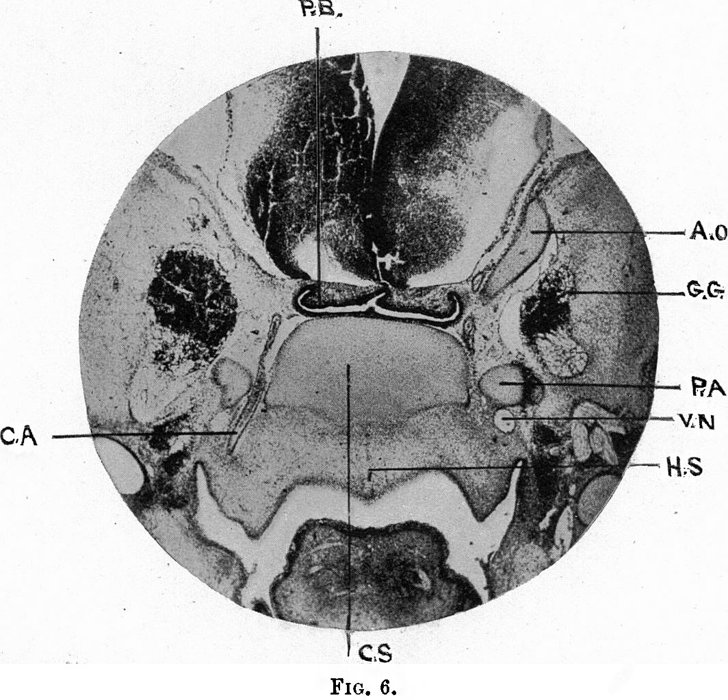 | 1,064 × 1,022 (285 KB) | Z8600021 (talk | contribs) | {{Fawcett1910_sphenoid_figures}} |
You cannot overwrite this file.
File usage
The following page uses this file: