File:Meyer1920 Plate 1.jpg: Difference between revisions
mNo edit summary |
mNo edit summary |
||
| (One intermediate revision by the same user not shown) | |||
| Line 1: | Line 1: | ||
==Plate 1== | ==Plate 1== | ||
<gallery> | |||
File:Meyer1920_fig01.jpg|Fig. 1. Cross-section of twin hydatiform chorionic vesicles within the tube. (Specimen No. 825.) X3. | |||
File:Meyer1920_fig02.jpg|Fig. 2. Hydatiform villi from same specimen in section. X 45. | |||
File:Meyer1920_fig03.jpg|Fig. 3. Embryo No. 1771, covered with magma. X 4. | |||
File:Meyer1920_fig04.jpg|Fig. 4. Cross-section of tube No. 1771. X 2. | |||
File:Meyer1920_fig05.jpg|Fig. 5. Cross-section of tube from same case. X 4. | |||
File:Meyer1920_fig06.jpg|Fig. 6. Hydatiform villi from same case. X 45. | |||
File:Meyer1920_fig07.jpg|Fig. 7. Hydatiform chorionic vesicle in loco with the tube incised. No. 2052. X 2. | |||
</gallery> | |||
[[:File:Meyer1920_Plate_1.jpg|Plate 1]]: [[:File:Meyer1920_fig01.jpg|Fig. 1]] | [[:File:Meyer1920_fig02.jpg|Fig. 2]] | [[:File:Meyer1920_fig03.jpg|Fig. 3]] | [[:File:Meyer1920_fig04.jpg|Fig. 4]] | [[:File:Meyer1920_fig05.jpg|Fig. 5]] | [[:File:Meyer1920_fig06.jpg|Fig. 6]] | [[:File:Meyer1920_fig07.jpg|Fig. 7]] | [[:File:Meyer1920_Plate_1.jpg|Plate 1]]: [[:File:Meyer1920_fig01.jpg|Fig. 1]] | [[:File:Meyer1920_fig02.jpg|Fig. 2]] | [[:File:Meyer1920_fig03.jpg|Fig. 3]] | [[:File:Meyer1920_fig04.jpg|Fig. 4]] | [[:File:Meyer1920_fig05.jpg|Fig. 5]] | [[:File:Meyer1920_fig06.jpg|Fig. 6]] | [[:File:Meyer1920_fig07.jpg|Fig. 7]] | ||
| Line 20: | Line 14: | ||
{{Meyer1920}} | {{Meyer1920}} | ||
[[Category:Hydatidiform Mole]] | |||
Latest revision as of 18:26, 12 May 2014
Plate 1
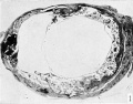
Fig. 1. Cross-section of twin hydatiform chorionic vesicles within the tube. (Specimen No. 825.) X3.
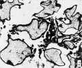
Fig. 2. Hydatiform villi from same specimen in section. X 45.
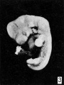
Fig. 3. Embryo No. 1771, covered with magma. X 4.
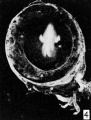
Fig. 4. Cross-section of tube No. 1771. X 2.
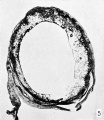
Fig. 5. Cross-section of tube from same case. X 4.
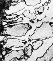
Fig. 6. Hydatiform villi from same case. X 45.
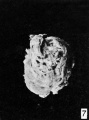
Fig. 7. Hydatiform chorionic vesicle in loco with the tube incised. No. 2052. X 2.


Plate 1: Fig. 1 | Fig. 2 | Fig. 3 | Fig. 4 | Fig. 5 | Fig. 6 | Fig. 7
- Meyer Links: Plate 1 | Plate 2 | Plate 3 | Plate 4 | Plate 5 | Plate 6 | Contribution No.40 | Volume IX | Contributions to Embryology | Hydatidiform Mole | Tubal Pregnancy
| Historic Disclaimer - information about historic embryology pages |
|---|
 |
Reference
Meyer AW. Hydatiform degeneration in tubal and uterine pregnancy. (1920) Carnegie Instn. Wash. Publ., Contrib. Embryol., 40: 327- 364.
Cite this page: Hill, M.A. (2024, June 26) Embryology Meyer1920 Plate 1.jpg. Retrieved from https://embryology.med.unsw.edu.au/embryology/index.php/File:Meyer1920_Plate_1.jpg
- © Dr Mark Hill 2024, UNSW Embryology ISBN: 978 0 7334 2609 4 - UNSW CRICOS Provider Code No. 00098G
File history
Yi efo/eka'e gwa ebo wo le nyangagi wuncin ye kamina wunga tinya nan
| Gwalagizhi | Nyangagi | Dimensions | User | Comment | |
|---|---|---|---|---|---|
| current | 18:57, 7 April 2012 | 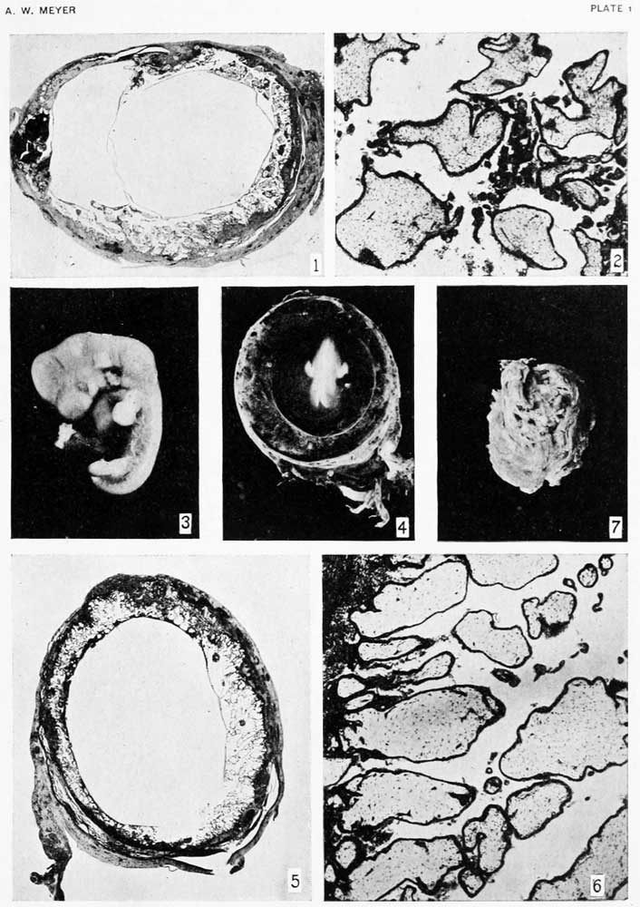 | 706 × 1,000 (113 KB) | Z8600021 (talk | contribs) | {{Meyer1920}} |
You cannot overwrite this file.
File usage
The following 2 pages use this file: