Category:Blue Histology: Difference between revisions
mNo edit summary |
mNo edit summary |
||
| (One intermediate revision by the same user not shown) | |||
| Line 1: | Line 1: | ||
{{External Links}} | |||
This {{Embryology}} category relates to [http://www.lab.anhb.uwa.edu.au/mb140/ Blue Histology] images copyright Lutz Slomianka 1998-2009. | This {{Embryology}} category relates to [http://www.lab.anhb.uwa.edu.au/mb140/ Blue Histology] images copyright Lutz Slomianka 1998-2009. | ||
Latest revision as of 14:05, 16 January 2015
External Links Notice - The dynamic nature of the internet may mean that some of these listed links may no longer function. If the link no longer works search the web with the link text or name. Links to any external commercial sites are provided for information purposes only and should never be considered an endorsement. UNSW Embryology is provided as an educational resource with no clinical information or commercial affiliation.
This Embryology category relates to Blue Histology images copyright Lutz Slomianka 1998-2009.
The literary and artistic works on the original Blue Histology website may be reproduced, adapted, published and distributed for non-commercial purposes.
Pages in category 'Blue Histology'
The following 5 pages are in this category, out of 5 total.
Media in category 'Blue Histology'
The following 200 files are in this category, out of 431 total.
(previous page) (next page)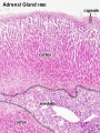 Adrenal histology 001.jpg 450 × 600; 151 KB
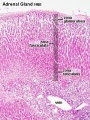 Adrenal histology 002.jpg 450 × 600; 150 KB
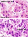 Adrenal histology 003.jpg 450 × 600; 71 KB
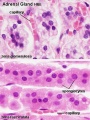 Adrenal histology 004.jpg 450 × 600; 71 KB
 Adrenal histology 005.jpg 1,280 × 1,024; 298 KB
 Adrenal histology 006.jpg 1,280 × 1,024; 313 KB
 Adrenal histology 007.jpg 1,280 × 1,024; 298 KB
 Adrenal histology 008.jpg 1,280 × 1,024; 272 KB
 Adrenal histology 009.jpg 1,280 × 1,024; 265 KB
 Adrenal histology 010.jpg 1,280 × 1,024; 292 KB
 Adrenal histology 011.jpg 1,280 × 1,024; 572 KB
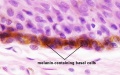
 Adult epidermis histology 01.jpg 600 × 750; 83 KB
 Adult epidermis histology 02.jpg 600 × 750; 123 KB
 Adult epidermis histology 03.jpg 600 × 375; 46 KB
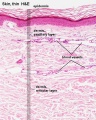 Adult skin histology 01.jpg 600 × 750; 73 KB
 Adult skin histology 02.jpg 600 × 750; 88 KB
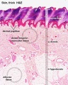
 Adult skin histology 03.jpg 600 × 750; 108 KB
 Adult skin histology 04.jpg 480 × 600; 128 KB
 Apocrine secretion animation.gif 60 × 80; 4 KB
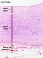
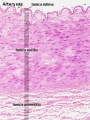 Artery histology 01.jpg 400 × 533; 80 KB
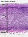
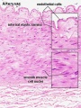 Artery histology 02.jpg 400 × 533; 78 KB
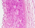
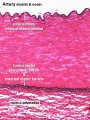 Artery histology 03.jpg 400 × 533; 89 KB
 Artery histology 04.jpg 800 × 1,000; 93 KB
 Artery histology 05.jpg 400 × 533; 72 KB
 Artery histology 06.jpg 400 × 533; 91 KB
 Artery histology 11.jpg 1,280 × 1,024; 344 KB
 Artery histology 12.jpg 1,280 × 1,024; 206 KB
 Artery histology 13.jpg 1,280 × 1,024; 474 KB
 Artery histology 14.jpg 1,280 × 1,024; 466 KB
 Artery histology 15.jpg 1,280 × 1,024; 343 KB
 Artery histology 16.jpg 1,280 × 1,024; 409 KB
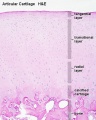
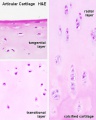 Articular cartilage 01.jpg 500 × 626; 42 KB
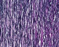
 Articular cartilage.jpg 500 × 626; 77 KB
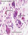 Autonomic ganglion histology 01.jpg 641 × 800; 56 KB
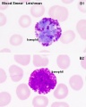 Basophil 01.jpg 480 × 600; 37 KB
 Bladder histology 001.jpg 1,280 × 1,024; 522 KB
 Bladder histology 002.jpg 1,280 × 1,024; 295 KB
 Bladder histology 003.jpg 1,280 × 1,024; 229 KB
 Bladder histology 004.jpg 1,280 × 1,024; 212 KB
 Bladder histology 01.jpg 480 × 600; 29 KB
 Bladder histology.jpg 300 × 400; 56 KB
 Blood vessel wall cartoon.jpg 450 × 600; 71 KB
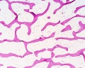
 Bone histology 001.jpg 1,280 × 1,024; 276 KB
 Bone histology 002.jpg 1,280 × 1,024; 309 KB
 Bone histology 003.jpg 1,280 × 1,024; 663 KB
 Bone histology 004.jpg 1,280 × 1,024; 605 KB
 Bone histology 005.jpg 1,280 × 1,024; 529 KB
 Bone histology 006.jpg 1,280 × 1,024; 360 KB
 Bone histology 007.jpg 1,280 × 1,024; 299 KB
 Bone histology 008.jpg 1,280 × 1,024; 550 KB
 Bone histology 009.jpg 1,280 × 1,024; 444 KB
 Bone histology 010.jpg 1,280 × 1,024; 256 KB
 Bone histology 011.jpg 1,280 × 1,024; 348 KB
 Bone histology 012.jpg 1,280 × 1,024; 165 KB
 Bone histology 013.jpg 1,280 × 1,024; 210 KB
 Bone histology 014.jpg 1,280 × 1,024; 541 KB
 Bone histology 015.jpg 1,280 × 1,024; 519 KB
 Bone histology 016.jpg 1,280 × 1,024; 379 KB
 Bone histology 017.jpg 1,280 × 1,024; 442 KB
 Bone histology 018.jpg 1,280 × 1,024; 336 KB
 Bone histology 019.jpg 1,280 × 1,024; 275 KB
 Bone histology 020.jpg 1,280 × 1,024; 272 KB
 Bone histology 021.jpg 1,280 × 1,024; 254 KB
 Bone histology 022.jpg 2,500 × 2,000; 328 KB
 Bone histology 066.jpg 2,500 × 2,000; 361 KB
 Bone histology 101.jpg 400 × 533; 59 KB
 Bone histology 111.jpg 400 × 533; 70 KB
 Bone histology 112.jpg 400 × 533; 46 KB
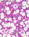
 Bone marrow histology 01.jpg 480 × 600; 114 KB
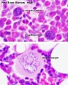
 Bone marrow histology 02.jpg 480 × 600; 109 KB
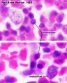
 Bone marrow histology 03.jpg 480 × 600; 81 KB
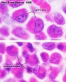
 Bone marrow histology 04.jpg 480 × 600; 61 KB
 Bone marrow histology 05.jpg 480 × 600; 62 KB
 Bone-bon02he.jpg 1,280 × 1,024; 348 KB
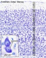 Brain histology 01.jpg 480 × 600; 125 KB
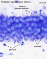 Brain histology 02.jpg 480 × 600; 51 KB
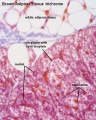 Brown adipose histology.jpg 400 × 500; 64 KB
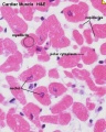
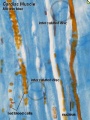 Cardiac muscle histology.jpg 300 × 400; 42 KB
 Cartilage em01.jpg 800 × 551; 176 KB
 Cartilage histology 001.jpg 1,280 × 1,024; 362 KB
 Cartilage histology 002.jpg 1,280 × 1,024; 174 KB
 Cartilage histology 003.jpg 1,280 × 1,024; 161 KB
 Cartilage histology 004.jpg 1,280 × 1,024; 216 KB
 Cartilage histology 005.jpg 1,280 × 1,024; 344 KB
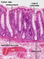
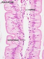
 Colon histology 001.jpg 400 × 533; 64 KB
 Colon histology 002.jpg 300 × 400; 73 KB
 Colon histology 003.jpg 1,280 × 1,024; 117 KB
 Colon histology 004.jpg 1,280 × 1,024; 292 KB
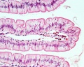
 Colon histology 006.jpg 400 × 533; 70 KB
 Colon histology 007.jpg 1,280 × 1,024; 152 KB
 Colon histology 008.jpg 1,278 × 959; 237 KB
 Colon histology 009.jpg 1,280 × 1,024; 159 KB
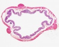
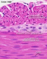
 Colon MALT.jpg 500 × 333; 67 KB
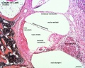
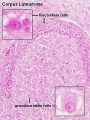 Corpus luteum lutein cells.jpg 450 × 600; 104 KB
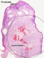 Corpus luteum.jpg 450 × 600; 94 KB
 Dorsal root ganglion histology 01.jpg 640 × 800; 45 KB
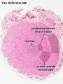 Ductus deferens 01.jpg 400 × 533; 76 KB
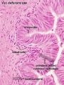 Ductus deferens 02.jpg 400 × 533; 80 KB
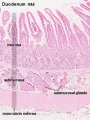
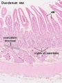
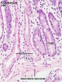
 Duodenum histology 01.jpg 480 × 600; 83 KB
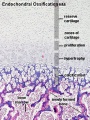
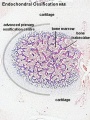 Endochondral ossification 1.jpg 400 × 534; 91 KB
 Endochondral ossification 2.jpg 400 × 533; 99 KB
 Endochondral ossification.jpg 400 × 533; 91 KB
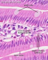
 Epididymis histology 01.jpg 600 × 375; 20 KB
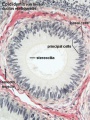 Epididymis histology 03.jpg 400 × 533; 68 KB
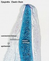 Epiglottis cartilage 01.jpg 500 × 626; 95 KB
 Epiglottis cartilage 02.jpg 500 × 626; 101 KB
 Fetal cartilage 01.jpg 639 × 400; 71 KB
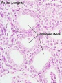
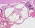
 Fetal lung histology 01.jpg 1,280 × 1,024; 339 KB
 Fetal lung histology 02.jpg 450 × 600; 74 KB
 Fetal lung histology.jpg 450 × 600; 83 KB
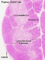 Fetal thymus.jpg 450 × 600; 122 KB
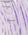 Fibrous cartilage 01.jpg 500 × 626; 76 KB
 Fibrous cartilage 02.jpg 500 × 626; 96 KB
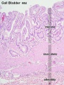 Gall bladder histology 001.jpg 375 × 500; 78 KB
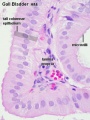 Gall bladder histology 002.jpg 375 × 500; 45 KB
 Gall bladder histology 003.jpg 1,280 × 1,024; 577 KB
 Gall bladder histology 004.jpg 1,280 × 1,024; 254 KB
 Gastrointestinal villi and crypts cartoon.jpg 500 × 333; 28 KB
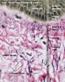
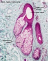
 Hair histology.jpg 600 × 451; 131 KB
 Heart histology 001.jpg 400 × 500; 83 KB
 Heart histology 002.jpg 400 × 500; 81 KB
 Heart histology 003.jpg 400 × 500; 136 KB
 Heart histology 004.jpg 400 × 500; 97 KB
 Heart histology 101.jpg 1,280 × 1,024; 258 KB
 Heart histology 102.jpg 1,280 × 1,024; 242 KB
 Heart histology 103.jpg 1,280 × 1,024; 281 KB
 Heart histology 104.jpg 1,280 × 1,024; 280 KB
 Heart histology 105.jpg 1,280 × 1,024; 379 KB
 Heart histology 106.jpg 1,280 × 1,024; 347 KB
 Heart histology 107.jpg 1,280 × 1,024; 395 KB
 Heart-histology-102.jpg 1,280 × 1,024; 242 KB
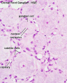
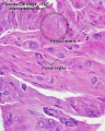
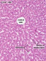
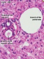
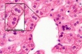
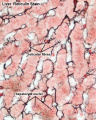
 Histology-fetal liver HEx100.jpg 1,280 × 1,024; 214 KB
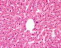
 Histology-fetal liver HEx40.jpg 1,000 × 800; 281 KB
 Holocrine secretion animation.gif 60 × 80; 16 KB
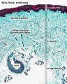
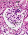
 Human fetal kidney histology 01.jpg 1,280 × 1,024; 481 KB
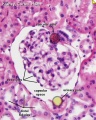
 Human fetal kidney histology 02.jpg 1,280 × 1,024; 322 KB
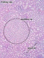
 Human fetal kidney histology 03.jpg 1,280 × 1,024; 333 KB
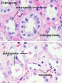
 Human fetal kidney histology 04.jpg 1,280 × 1,024; 307 KB
 Hyaline cartilage 01.jpg 500 × 626; 71 KB
 Hyaline cartilage 02.jpg 500 × 626; 62 KB
 Hyaline cartilage 03.jpg 500 × 626; 92 KB
 Hyaline cartilage 04.jpg 500 × 626; 101 KB
 Ileum histology 01.jpg 480 × 600; 69 KB
 Integumentary histology 01.jpg 480 × 600; 70 KB
 Integumentary histology 04.jpg 280 × 700; 65 KB
 Integumentary histology 10.jpg 800 × 1,000; 101 KB
 Integumentary- hair follicle 01.jpg 479 × 599; 66 KB
 Integumentary- hair follicle 02.jpg 479 × 599; 74 KB
 Integumentary- hair follicle 03.jpg 479 × 599; 48 KB
 Integumentary- sebaceous gland histology 01.jpg 400 × 500; 125 KB
 Integumentary- sebaceous gland histology 02.jpg 400 × 500; 150 KB
 Intestine histology 001.jpg 450 × 600; 65 KB
 Intestine histology 002.jpg 800 × 640; 130 KB
 Intestine histology 003.jpg 400 × 533; 64 KB
 Intestine histology 004.jpg 400 × 533; 81 KB
 Intestine histology 005.jpg 400 × 533; 78 KB
 Intestine histology 006.jpg 400 × 533; 77 KB
 Intestine histology 007.jpg 400 × 533; 82 KB
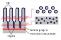 Intestine villi crypts cartoon.jpg 500 × 334; 29 KB
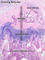
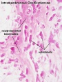 Intramembranous ossification centre.jpg 450 × 600; 69 KB
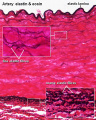
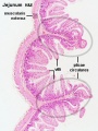
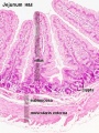
 Jejunum histology 01.jpg 480 × 600; 79 KB
 Liver animated cartoon.gif 300 × 200; 239 KB
 Liver histology 001.jpg 400 × 533; 94 KB
 Liver histology 002.jpg 375 × 500; 54 KB
 Liver histology 003.jpg 375 × 500; 52 KB
 Liver histology 004.jpg 600 × 400; 70 KB
 Liver histology 008.jpg 1,280 × 1,024; 214 KB
 Liver histology 009.jpg 1,280 × 1,024; 373 KB
 Liver histology 101.jpg 1,280 × 1,024; 410 KB
 Liver histology 102.jpg 1,280 × 1,024; 475 KB
 Liver histology 103.jpg 1,280 × 1,024; 330 KB
 Liver histology EM01.jpg 1,028 × 708; 141 KB
 Liver histology EM02.jpg 1,028 × 707; 154 KB
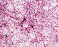
 Liver- Kupffer cell and reticular fibre.jpg 600 × 800; 49 KB
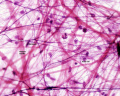
 Liver-reticular fibre.jpg 700 × 875; 77 KB
 Lymph node 05.jpg 1,000 × 800; 180 KB
 Lymph node histology 01.jpg 600 × 400; 61 KB
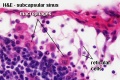
 Lymph node histology 02.jpg 450 × 600; 130 KB
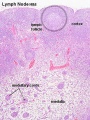
 Lymph node histology 03.jpg 450 × 600; 140 KB
 Lymph node histology 04.jpg 450 × 600; 88 KB
 Lymph node histology 05.jpg 450 × 600; 87 KB
 Lymph node histology 06.jpg 450 × 600; 141 KB
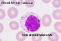 Lymphocyte 02.jpg 500 × 334; 27 KB
 Merocrine secretion animation.gif 60 × 80; 10 KB
 Mesentery histology 01.jpg 1,280 × 1,024; 139 KB
 Mesentery histology 02.jpg 1,280 × 1,024; 256 KB
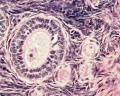 Monkey- ovary primordial follicle.jpg 1,000 × 800; 292 KB
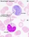 Monocyte 01.jpg 480 × 600; 39 KB
 Mouse organ of corti 01.jpg 1,280 × 1,024; 339 KB
 Mouse organ of corti 02.jpg 1,280 × 1,024; 320 KB
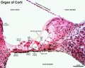 Mouse organ of corti 03.jpg 1,280 × 1,024; 207 KB
 Mouse organ of corti 04.jpg 1,280 × 1,024; 202 KB
 Mouse organ of corti 05.jpg 1,280 × 1,024; 171 KB
 Muscle fiber types.jpg 400 × 250; 49 KB
 Myelination animation.gif 300 × 200; 77 KB
 Nephron histology 01.jpg 400 × 500; 79 KB
 Nephron histology 02.jpg 400 × 500; 77 KB
 Nephron histology 03.jpg 375 × 500; 97 KB
 Nephron histology 04.jpg 375 × 500; 54 KB
 Nephron histology.jpg 400 × 500; 70 KB


