File:Cardiac muscle EM01.jpg: Difference between revisions
| Line 1: | Line 1: | ||
==Cardiac Muscle Electron Micrograph== | ==Cardiac Muscle Electron Micrograph== | ||
Electron micrograph of parts of three cat cardiac muscle fibers and an adjacent capillary in longitudinal section. | Electron micrograph of parts of three cat cardiac muscle fibers and an adjacent capillary in longitudinal section. This is a historic (1969) EM showing key features in cardiac muscle ultrastructure. Only the intercalated disc and some cross-striations can typically be seen in the light microscope histology slides. | ||
* '''In D''' - Intercalated disc, the two upper cells are joined end to end by a typical steplike intercalated disc. | * '''In D''' - Intercalated disc, the two upper cells are joined end to end by a typical steplike intercalated disc. | ||
Revision as of 15:01, 6 August 2012
Cardiac Muscle Electron Micrograph
Electron micrograph of parts of three cat cardiac muscle fibers and an adjacent capillary in longitudinal section. This is a historic (1969) EM showing key features in cardiac muscle ultrastructure. Only the intercalated disc and some cross-striations can typically be seen in the light microscope histology slides.
- In D - Intercalated disc, the two upper cells are joined end to end by a typical steplike intercalated disc.
- Mt - Rows of mitochondria appear to divide the contractile substance into myofibril-like units but, unlike the true myofibrils of skeletal muscle, these branch and rejoin and are quite variable in width.
- Lp - Lipid droplets somewhat distorted in specimen preparation are found between the ends of the mitochondria.
- Cap - Capillary.
Original image X 15,000.
Reference
<pubmed>4891913</pubmed>| PMC2107571
Copyright
Rockefeller University Press - Copyright Policy This article is distributed under the terms of an Attribution–Noncommercial–Share Alike–No Mirror Sites license for the first six months after the publication date (see http://www.jcb.org/misc/terms.shtml). After six months it is available under a Creative Commons License (Attribution–Noncommercial–Share Alike 4.0 Unported license, as described at https://creativecommons.org/licenses/by-nc-sa/4.0/ ). (More? Help:Copyright Tutorial)
File history
Yi efo/eka'e gwa ebo wo le nyangagi wuncin ye kamina wunga tinya nan
| Gwalagizhi | Nyangagi | Dimensions | User | Comment | |
|---|---|---|---|---|---|
| current | 12:52, 6 August 2012 | 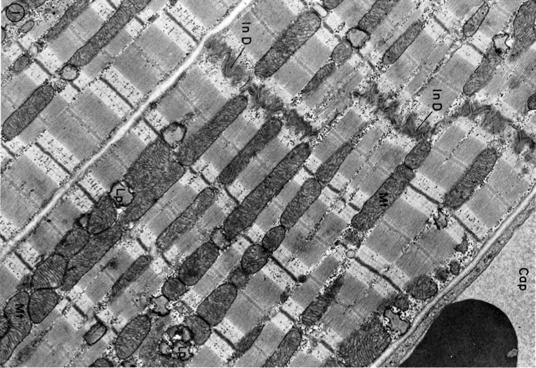 | 1,072 × 735 (231 KB) | Z8600021 (talk | contribs) | ==Cardiac Muscle EM== Electron micrograph of parts of three cat cardiac muscle fibers and an adjacent capillary (Cap) in longitudinal section. The two upper cells are joined end to end by a typical steplike intercalated disc (In D).Rows of mitochondria ( |
You cannot overwrite this file.
File usage
The following 5 pages use this file:


