File:Mouse oocyte cortical granules 01.jpg: Difference between revisions
(==Mouse oocyte cortical granules== Confocal scanning laser micrographs of cortical granules in mouse oocytes labeled with lectin LCA. (A) An equatorial section of unfertilized zona free oocyte and (B) a three-dimensional projection of unfertilized zona i) |
(No difference)
|
Revision as of 15:57, 6 May 2012
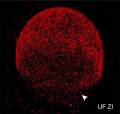
Mouse oocyte cortical granules
Confocal scanning laser micrographs of cortical granules in mouse oocytes labeled with lectin LCA. (A) An equatorial section of unfertilized zona free oocyte and (B) a three-dimensional projection of unfertilized zona intact oocyte showing the cortical granules (in red), the second cortical granule free domain (arrows), and the pre-fertilization release (arrowhead).
Reference
http://www.rbej.com/content/9/1/149
© 2011 Liu; licensee BioMed Central Ltd.
This is an Open Access article distributed under the terms of the Creative Commons Attribution License (http://creativecommons.org/licenses/by/2.0), which permits unrestricted use, distribution, and reproduction in any medium, provided the original work is properly cited.
Panel B cropped and resized from Figure 2. Original file name 1477-7827-9-149-2.jpg
File history
Yi efo/eka'e gwa ebo wo le nyangagi wuncin ye kamina wunga tinya nan
| Gwalagizhi | Nyangagi | Dimensions | User | Comment | |
|---|---|---|---|---|---|
| current | 15:57, 6 May 2012 |  | 500 × 475 (53 KB) | Z8600021 (talk | contribs) | ==Mouse oocyte cortical granules== Confocal scanning laser micrographs of cortical granules in mouse oocytes labeled with lectin LCA. (A) An equatorial section of unfertilized zona free oocyte and (B) a three-dimensional projection of unfertilized zona i |
You cannot overwrite this file.
File usage
The following 2 pages use this file: