File:Skull CT abnormal 02.jpg: Difference between revisions
No edit summary |
No edit summary |
||
| Line 1: | Line 1: | ||
==Skull Coronal Synostosis== | |||
There is complete fusion of the coronal suture (white arrows) with a prominent frontal bone and flattened occiput. Coronal reconstruction (D) demonstrates prominent bilateral elliptical orbits, known as the "harlequin eye" deformity. Note the early partial fusion of the right coronal suture (arrowheads in E) | |||
* '''A-C''' - Bilateral coronal synostosis 3DCT volume rendered images. | |||
* '''D''' - Bilateral coronal synostosis, coronal CT scan. | |||
* '''E''' - unilateral partial coronal synostosis 3DCT volume rendered images | |||
===Reference=== | ===Reference=== | ||
<pubmed>21431034</pubmed>| [http://www.ncbi.nlm.nih.gov/pmc/articles/PMC3056371 PMC3056371] | [http://www.ijri.org/article.asp?issn=0971-3026;year=2011;volume=21;issue=1;spage=49;epage=56;aulast=Khanna Indian J Radiol Imaging.] | <pubmed>21431034</pubmed>| [http://www.ncbi.nlm.nih.gov/pmc/articles/PMC3056371 PMC3056371] | [http://www.ijri.org/article.asp?issn=0971-3026;year=2011;volume=21;issue=1;spage=49;epage=56;aulast=Khanna Indian J Radiol Imaging.] | ||
| Line 10: | Line 19: | ||
Original file name: Original figure has been modified, resized and relabelled. | Original file name: Figure 3 (A-E) Original figure has been modified, resized and relabelled. | ||
http://www.ijri.org/viewimage.asp?img=IndianJRadiolImaging_2011_21_1_49_76055_f4.jpg | |||
[[Category:Human]] [[Category:Skull]] [[Category:Abnormal]] [[Category:Computed Tomography]] | [[Category:Human]] [[Category:Skull]] [[Category:Abnormal Development]] [[Category:Computed Tomography]] | ||
Revision as of 11:22, 17 March 2012
Skull Coronal Synostosis
There is complete fusion of the coronal suture (white arrows) with a prominent frontal bone and flattened occiput. Coronal reconstruction (D) demonstrates prominent bilateral elliptical orbits, known as the "harlequin eye" deformity. Note the early partial fusion of the right coronal suture (arrowheads in E)
- A-C - Bilateral coronal synostosis 3DCT volume rendered images.
- D - Bilateral coronal synostosis, coronal CT scan.
- E - unilateral partial coronal synostosis 3DCT volume rendered images
Reference
<pubmed>21431034</pubmed>| PMC3056371 | Indian J Radiol Imaging.
This is an open-access article distributed under the terms of the Creative Commons Attribution License, which permits unrestricted use, distribution, and reproduction in any medium, provided the original work is properly cited.
Paritosh C Khanna © 2007 - 2012 Indian Journal of Radiology and Imaging
Attribution-NonCommercial-ShareAlike 3.0 Unported (CC BY-NC-SA 3.0)
Original file name: Figure 3 (A-E) Original figure has been modified, resized and relabelled.
http://www.ijri.org/viewimage.asp?img=IndianJRadiolImaging_2011_21_1_49_76055_f4.jpg
File history
Yi efo/eka'e gwa ebo wo le nyangagi wuncin ye kamina wunga tinya nan
| Gwalagizhi | Nyangagi | Dimensions | User | Comment | |
|---|---|---|---|---|---|
| current | 11:10, 17 March 2012 | 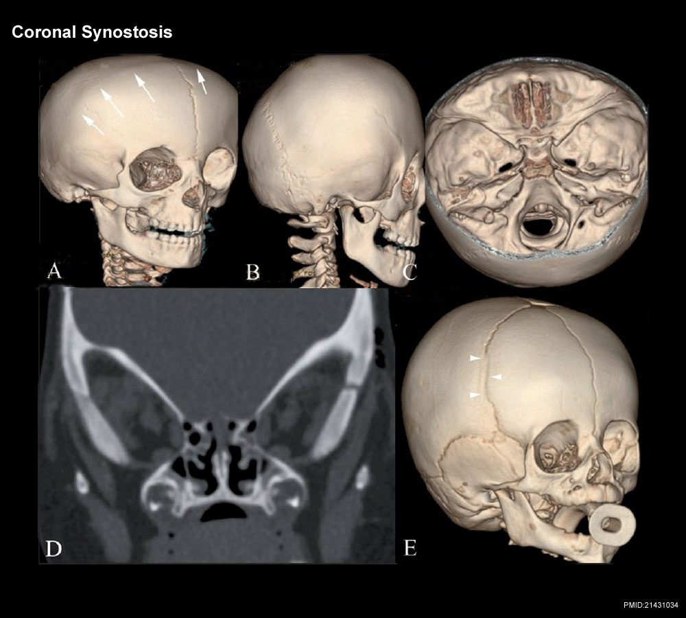 | 1,000 × 900 (119 KB) | Z8600021 (talk | contribs) |
You cannot overwrite this file.
File usage
The following 4 pages use this file: