File:T2 lymphocyte EM13.jpg: Difference between revisions
From Embryology
(==T2 lymphocyte Electron Micrograph== FIg. 13. Spleen cell (10 days after alloantigenic immunization with DBA/2 mastocytoma) labeled with aMSLA detected by the bridge technique with phage T4. Some of the phage heads are sectioned tangentially and theref) |
(No difference)
|
Revision as of 16:23, 22 February 2012
T2 lymphocyte Electron Micrograph
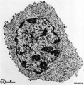
FIg. 13. Spleen cell (10 days after alloantigenic immunization with DBA/2 mastocytoma) labeled with aMSLA detected by the bridge technique with phage T4. Some of the phage heads are sectioned tangentially and therefore barely visible.
This large blast-like cell, classified as T2 lymphocyte, is mostly characterized by its very large content in polyribosomes. X 15,000.
File history
Yi efo/eka'e gwa ebo wo le nyangagi wuncin ye kamina wunga tinya nan
| Gwalagizhi | Nyangagi | Dimensions | User | Comment | |
|---|---|---|---|---|---|
| current | 16:23, 22 February 2012 |  | 781 × 795 (177 KB) | Z8600021 (talk | contribs) | ==T2 lymphocyte Electron Micrograph== FIg. 13. Spleen cell (10 days after alloantigenic immunization with DBA/2 mastocytoma) labeled with aMSLA detected by the bridge technique with phage T4. Some of the phage heads are sectioned tangentially and theref |
You cannot overwrite this file.
File usage
The following 8 pages use this file: