File:Aortic arch and ductus arteriosus.jpg: Difference between revisions
No edit summary |
|||
| (3 intermediate revisions by the same user not shown) | |||
| Line 9: | Line 9: | ||
(c) In aortic coarctation, a left sided heart malformation, the DA is kept open by administration of PGE allowing blood to enter the systemic circulation. Dashed lines indicate the plane of tissue sections shown in d–h. | (c) In aortic coarctation, a left sided heart malformation, the DA is kept open by administration of PGE allowing blood to enter the systemic circulation. Dashed lines indicate the plane of tissue sections shown in d–h. | ||
(d;1 in c) Transverse sections of the muscular DA, | |||
(e;2 in a) the elastic aorta | |||
(f;3 in b) the ligamentum arteriosum. Intimal thickening in the DA is indicated in yellow. | |||
Areas with CN are indicated in white and the wavy elastic lamellae with blue. Endothelial cells (red outlines) line the inside of the vessels. Longitudinal sections through the DA (4 in a, providing Fig. 1g, and 5 in c providing Fig. 1h). | |||
(g) DA tissue and aorta tissue are merging and can be separately distinguished by their characteristic histology. | (g) DA tissue and aorta tissue are merging and can be separately distinguished by their characteristic histology. | ||
(h) In coarctation, inner media and intimal thickening of the DA extend into the aorta sometimes almost encircling the lumen of the aortic arch at that site (as depicted). The aortic tissue will always be present in the roof of the coarctation. | (h) In coarctation, inner media and intimal thickening of the DA extend into the aorta sometimes almost encircling the lumen of the aortic arch at that site (as depicted). The aortic tissue will always be present in the roof of the coarctation. | ||
:'''Links:''' [[Cardiovascular System Development]] | |||
| Line 19: | Line 28: | ||
===Reference=== | ===Reference=== | ||
<pubmed>21915271</pubmed>| [http://www.plosone.org/article/info%3Adoi%2F10.1371%2Fjournal.pone.0023975 PLoS One.] | |||
http://www.plosone.org/article/info%3Adoi%2F10.1371%2Fjournal.pone.0023975 | |||
© 2011 Bokenkamp et al. This is an open-access article distributed under the terms of the Creative Commons Attribution License, which permits unrestricted use, distribution, and reproduction in any medium, provided the original author and source are credited. | © 2011 Bokenkamp et al. This is an open-access article distributed under the terms of the Creative Commons Attribution License, which permits unrestricted use, distribution, and reproduction in any medium, provided the original author and source are credited. | ||
[[Category:Cardiovascular]] [[Category:Heart]] | [[Category:Cardiovascular]] [[Category:Heart]] [[Category:Cartoon]] [[Category:Arterial]] | ||
Latest revision as of 12:45, 20 October 2011
Schematic presentations of the great vessels
Anatomy of DA and the adjacent arteries.
(a) A patent DA is found in the fetus before respiration starts. Oxygen-rich blood (in red) from the placenta passes though the aorta. Oxygen-poor blood from the fetus (in blue) passes the DA entering the descending aorta (in purple).
(b) After the start of respiration the DA regresses into a ligamentum arteriosum. Aortic (red) and pulmonary (blue) circulation are disconnected.
(c) In aortic coarctation, a left sided heart malformation, the DA is kept open by administration of PGE allowing blood to enter the systemic circulation. Dashed lines indicate the plane of tissue sections shown in d–h.
(d;1 in c) Transverse sections of the muscular DA,
(e;2 in a) the elastic aorta
(f;3 in b) the ligamentum arteriosum. Intimal thickening in the DA is indicated in yellow.
Areas with CN are indicated in white and the wavy elastic lamellae with blue. Endothelial cells (red outlines) line the inside of the vessels. Longitudinal sections through the DA (4 in a, providing Fig. 1g, and 5 in c providing Fig. 1h).
(g) DA tissue and aorta tissue are merging and can be separately distinguished by their characteristic histology.
(h) In coarctation, inner media and intimal thickening of the DA extend into the aorta sometimes almost encircling the lumen of the aortic arch at that site (as depicted). The aortic tissue will always be present in the roof of the coarctation.
Figure 1. Journal.pone.0023975.g001.jpg doi:10.1371/journal.pone.0023975.g001
Reference
<pubmed>21915271</pubmed>| PLoS One.
© 2011 Bokenkamp et al. This is an open-access article distributed under the terms of the Creative Commons Attribution License, which permits unrestricted use, distribution, and reproduction in any medium, provided the original author and source are credited.
File history
Yi efo/eka'e gwa ebo wo le nyangagi wuncin ye kamina wunga tinya nan
| Gwalagizhi | Nyangagi | Dimensions | User | Comment | |
|---|---|---|---|---|---|
| current | 22:15, 7 September 2011 | 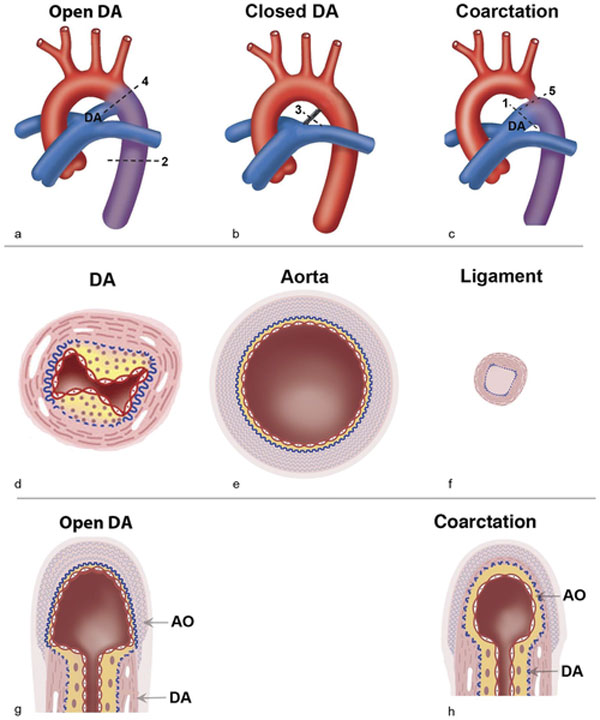 | 600 × 720 (68 KB) | S8600021 (talk | contribs) | ==Schematic presentations of the great vessels== Anatomy of DA and the adjacent arteries. (a) A patent DA is found in the fetus before respiration starts. Oxygen-rich blood (in red) from the placenta passes though the aorta. Oxygen-poor blood from the |
You cannot overwrite this file.
File usage
There are no pages that use this file.