File:Kollmann354.jpg: Difference between revisions
({{Kollmann1907}}) |
No edit summary |
||
| Line 1: | Line 1: | ||
==Fig. 354. Sagittal section through the head of a human embryo CRL of 4.2 mm, 31 - 34 days old== | |||
(After His.) | |||
The right wall of the foregut is exposed to the four internal gill | |||
bags of decreasing size from top to bottom. This part of the foregut | |||
located directly behind the heart. At the entrance of the mouth: the upper and | |||
extension of the lower jaw, some dorsal raises the Rathke's pouch | |||
behind her the head end of the notochord is that of the ventral | |||
Caudal neural tube pulls, trapped in the mesoderm of the primitive | |||
Spinal column. Below the last inner branchial pouch develops from the | |||
the space of the ventral foregut, the lung unit in the form of a trough, | |||
Lung channel, pulmonary sulcus, which some of its cranial end of- | |||
widened, which is simultaneously the separation be-in dining and trachea | |||
is derived. The channel ends as a blind sac-like "unpaired lung sacs" (Sacculus pulmonalis impar). | |||
{{Kollmann1907}} | {{Kollmann1907}} | ||
Fig. 354. Sagittalschnitt durch den Kopf eines menschlichen Embryo | |||
von 4,2 mm Nackensteißlänge, 31 — 34 Tage alt. | |||
(Nach His.) | |||
Die rechte Wand des Kopfdarms liegt frei mit den 4 inneren Kiemen- | |||
taschen von oben nach unten an Größe abnehmend. Dieser Teil des Kopfdarms | |||
liegt direkt hinter dem Herzen. An dem Eingang des Mundes: der Ober- und | |||
der Unterkieferfortsatz, etwas dorsal erhebt sich die Rathkesche Tasche, | |||
hinter ihr liegt das Kopfende der Chorda dorsalis, welche ventral von dem | |||
MeduUarrohr kaudalwärts zieht, eingeschlossen in das Mesoderm der primitiven | |||
Wirbelsäule. Unterhalb der letzten inneren Kiementasche entwickelt sich aus | |||
dem Raum des Kopfdarms ventral die Lungenanlage in Form einer Rinne, | |||
Lungenrinne, Sulcus pulmonalis, welche an ihrem kranialen Ende etwas aus- | |||
geweitet ist, wobei gleichzeitig die Sonderung in Speise- und Luftröhre einge- | |||
leitet wird. Die Rinne endigt blindsackförmig als „unpaares Lungensäckchen", | |||
Sacculus pulmonalis impar. | |||
Latest revision as of 15:36, 17 October 2011
Fig. 354. Sagittal section through the head of a human embryo CRL of 4.2 mm, 31 - 34 days old
(After His.)
The right wall of the foregut is exposed to the four internal gill
bags of decreasing size from top to bottom. This part of the foregut
located directly behind the heart. At the entrance of the mouth: the upper and
extension of the lower jaw, some dorsal raises the Rathke's pouch
behind her the head end of the notochord is that of the ventral
Caudal neural tube pulls, trapped in the mesoderm of the primitive
Spinal column. Below the last inner branchial pouch develops from the
the space of the ventral foregut, the lung unit in the form of a trough,
Lung channel, pulmonary sulcus, which some of its cranial end of-
widened, which is simultaneously the separation be-in dining and trachea
is derived. The channel ends as a blind sac-like "unpaired lung sacs" (Sacculus pulmonalis impar).
- This text is a Google translate computer generated translation and may contain many errors.
Images from - Atlas of the Development of Man (Volume 2)
(Handatlas der entwicklungsgeschichte des menschen)
- Kollmann Atlas 2: Gastrointestinal | Respiratory | Urogenital | Cardiovascular | Neural | Integumentary | Smell | Vision | Hearing | Kollmann Atlas 1 | Kollmann Atlas 2 | Julius Kollmann
- Links: Julius Kollman | Atlas Vol.1 | Atlas Vol.2 | Embryology History
| Historic Disclaimer - information about historic embryology pages |
|---|
 |
Reference
Kollmann JKE. Atlas of the Development of Man (Handatlas der entwicklungsgeschichte des menschen). (1907) Vol.1 and Vol. 2. Jena, Gustav Fischer. (1898).
Cite this page: Hill, M.A. (2024, June 26) Embryology Kollmann354.jpg. Retrieved from https://embryology.med.unsw.edu.au/embryology/index.php/File:Kollmann354.jpg
- © Dr Mark Hill 2024, UNSW Embryology ISBN: 978 0 7334 2609 4 - UNSW CRICOS Provider Code No. 00098G
Fig. 354. Sagittalschnitt durch den Kopf eines menschlichen Embryo
von 4,2 mm Nackensteißlänge, 31 — 34 Tage alt.
(Nach His.)
Die rechte Wand des Kopfdarms liegt frei mit den 4 inneren Kiemen- taschen von oben nach unten an Größe abnehmend. Dieser Teil des Kopfdarms liegt direkt hinter dem Herzen. An dem Eingang des Mundes: der Ober- und der Unterkieferfortsatz, etwas dorsal erhebt sich die Rathkesche Tasche, hinter ihr liegt das Kopfende der Chorda dorsalis, welche ventral von dem MeduUarrohr kaudalwärts zieht, eingeschlossen in das Mesoderm der primitiven Wirbelsäule. Unterhalb der letzten inneren Kiementasche entwickelt sich aus dem Raum des Kopfdarms ventral die Lungenanlage in Form einer Rinne, Lungenrinne, Sulcus pulmonalis, welche an ihrem kranialen Ende etwas aus- geweitet ist, wobei gleichzeitig die Sonderung in Speise- und Luftröhre einge- leitet wird. Die Rinne endigt blindsackförmig als „unpaares Lungensäckchen", Sacculus pulmonalis impar.
File history
Yi efo/eka'e gwa ebo wo le nyangagi wuncin ye kamina wunga tinya nan
| Gwalagizhi | Nyangagi | Dimensions | User | Comment | |
|---|---|---|---|---|---|
| current | 12:59, 16 October 2011 | 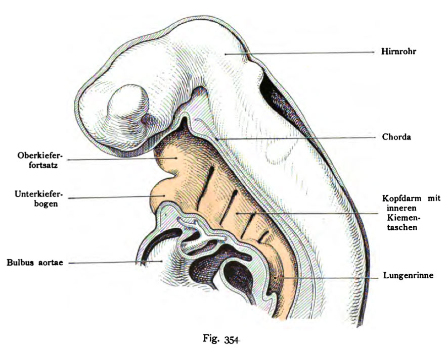 | 884 × 697 (101 KB) | S8600021 (talk | contribs) | {{Kollmann1907}} |
You cannot overwrite this file.
File usage
The following page uses this file: