File:Mitochondria EM01.jpg: Difference between revisions
From Embryology
(==Mitochondria Electron Micrograph== Transmission electron microscope image of a thin section cut through an area of mammalian lung tissue. The high magnification image shows a mitochondria. JEOL 100CX TEM Writer: Louisa Howard Keywords: mitochondria,) |
No edit summary |
||
| Line 14: | Line 14: | ||
[[Category:Mitochondria]] [[Category: | [[Category:Mitochondria]] [[Category:Electron Micrograph]] | ||
Revision as of 23:54, 22 September 2011
Mitochondria Electron Micrograph
Transmission electron microscope image of a thin section cut through an area of mammalian lung tissue. The high magnification image shows a mitochondria.
JEOL 100CX TEM
Writer: Louisa Howard
Keywords: mitochondria, mammalian, TEM, lung
Image Source: Dartmouth EM images, images are in the public domain.
File history
Yi efo/eka'e gwa ebo wo le nyangagi wuncin ye kamina wunga tinya nan
| Gwalagizhi | Nyangagi | Dimensions | User | Comment | |
|---|---|---|---|---|---|
| current | 11:47, 23 Haziran 2011 | 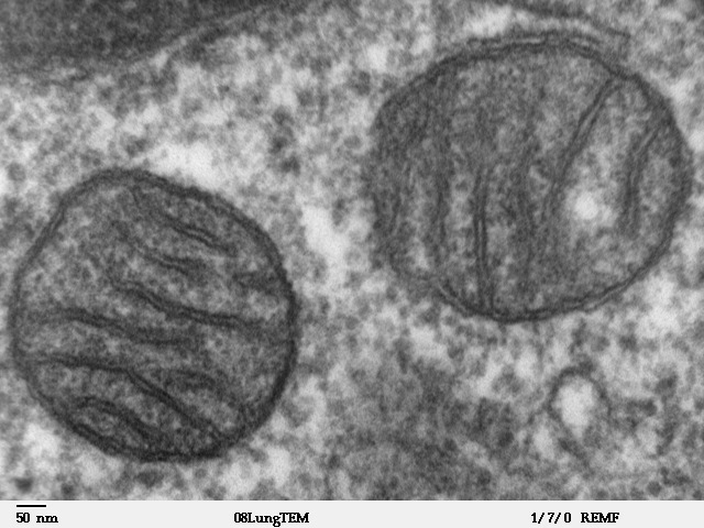 | 640 × 480 (96 KB) | S8600021 (talk | contribs) | ==Mitochondria Electron Micrograph== Transmission electron microscope image of a thin section cut through an area of mammalian lung tissue. The high magnification image shows a mitochondria. JEOL 100CX TEM Writer: Louisa Howard Keywords: mitochondria, |
You cannot overwrite this file.
File usage
The following 3 pages use this file:


