File:Ureter histology 001.jpg: Difference between revisions
From Embryology
(uploaded a new version of "File:Ureter histology 001.jpg") |
No edit summary |
||
| Line 1: | Line 1: | ||
==Ureter Histology== | |||
Ure10he.jpg | Ure10he.jpg | ||
{{Renal Histology}} | |||
{{Blue Histology}} | |||
Latest revision as of 18:05, 8 July 2011
Ureter Histology
Ure10he.jpg
- Renal System Histology: Nephron tubule overview | glomerulus structure | vascular and renal poles | Medullary rays | Nephron tubules
Links: Histology | Histology Stains | Blue Histology images copyright Lutz Slomianka 1998-2009. The literary and artistic works on the original Blue Histology website may be reproduced, adapted, published and distributed for non-commercial purposes. See also the page Histology Stains.
Cite this page: Hill, M.A. (2024, June 27) Embryology Ureter histology 001.jpg. Retrieved from https://embryology.med.unsw.edu.au/embryology/index.php/File:Ureter_histology_001.jpg
- © Dr Mark Hill 2024, UNSW Embryology ISBN: 978 0 7334 2609 4 - UNSW CRICOS Provider Code No. 00098G
File history
Yi efo/eka'e gwa ebo wo le nyangagi wuncin ye kamina wunga tinya nan
| Gwalagizhi | Nyangagi | Dimensions | User | Comment | |
|---|---|---|---|---|---|
| current | 15:11, 8 July 2011 | 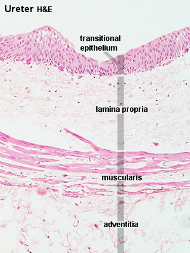 | 375 × 500 (50 KB) | S8600021 (talk | contribs) | |
| 15:09, 8 July 2011 | 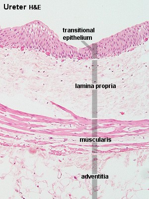 | 300 × 400 (45 KB) | S8600021 (talk | contribs) | Ure10he.jpg |
You cannot overwrite this file.
File usage
The following 5 pages use this file: