File:Ingalls1932b fig10.jpg: Difference between revisions
From Embryology
(==Plate 101== Fig. 9. Embryo No. 652. Greatest length 15 mm. Dorsal view of anterior end of embryo to show the transverse discolored band just behind the midbrain. Fig. 10. Embryo No. 167. Greatest length 18.5mm. Very conspicuous, sharply defined, discolored area over the rhombencephalon. There is a similar smaller patch over the vertex and two paired spots on the upper part of the forehead. Fig. 11. Embryo No. 671. Greatest length 25 mm. Enormous blood-stained bleb on back of head and nec...) |
mNo edit summary |
||
| Line 1: | Line 1: | ||
== | ==Fig. 10. Embryo No. 167== | ||
Greatest length 18.5mm. Very conspicuous, sharply defined, discolored area over the rhombencephalon. There is a similar smaller patch over the vertex and two paired spots on the upper part of the forehead. | |||
Latest revision as of 09:34, 14 October 2020
Fig. 10. Embryo No. 167
Greatest length 18.5mm. Very conspicuous, sharply defined, discolored area over the rhombencephalon. There is a similar smaller patch over the vertex and two paired spots on the upper part of the forehead.
Reference
Ingalls NW. Studies in the pathology of development: II. Some aspects of defective development in the dorsal midline. (1932) Am J Pathol. 8(5): 525-556 PMID 19970035
Cite this page: Hill, M.A. (2024, June 27) Embryology Ingalls1932b fig10.jpg. Retrieved from https://embryology.med.unsw.edu.au/embryology/index.php/File:Ingalls1932b_fig10.jpg
- © Dr Mark Hill 2024, UNSW Embryology ISBN: 978 0 7334 2609 4 - UNSW CRICOS Provider Code No. 00098G
File history
Yi efo/eka'e gwa ebo wo le nyangagi wuncin ye kamina wunga tinya nan
| Gwalagizhi | Nyangagi | Dimensions | User | Comment | |
|---|---|---|---|---|---|
| current | 09:32, 14 October 2020 | 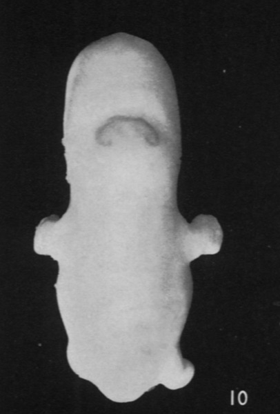 | 406 × 600 (41 KB) | Z8600021 (talk | contribs) | ==Plate 101== Fig. 9. Embryo No. 652. Greatest length 15 mm. Dorsal view of anterior end of embryo to show the transverse discolored band just behind the midbrain. Fig. 10. Embryo No. 167. Greatest length 18.5mm. Very conspicuous, sharply defined, discolored area over the rhombencephalon. There is a similar smaller patch over the vertex and two paired spots on the upper part of the forehead. Fig. 11. Embryo No. 671. Greatest length 25 mm. Enormous blood-stained bleb on back of head and nec... |
You cannot overwrite this file.
File usage
The following 2 pages use this file: