File:Epidermis cartoon 02.jpg: Difference between revisions
No edit summary |
|||
| Line 10: | Line 10: | ||
<pubmed>18209104</pubmed>[http://jcb.rupress.org/content/180/2/273.long JCB] | <pubmed>18209104</pubmed>[http://jcb.rupress.org/content/180/2/273.long JCB] | ||
[[Category:Integumentary]] [[Category:Cartoon]] | |||
{{Template:JCB}} | {{Template:JCB}} | ||
Revision as of 12:39, 13 October 2010
Epidermal Differentiation
The program of epidermal differentiation is shown in this schematic, illustrating the basement membrane at the base, the proliferative basal layer, and the three differentiation stages: spinous layer, granular layer, and outermost stratum corneum.
Related Image: same image with layer molecular information
Original file name: Figure 1. http://jcb.rupress.org/content/180/2/273/F1.expansion.html (resized and molecular information cropped)
Reference
<pubmed>18209104</pubmed>JCB
Copyright
Rockefeller University Press - Copyright Policy This article is distributed under the terms of an Attribution–Noncommercial–Share Alike–No Mirror Sites license for the first six months after the publication date (see http://www.jcb.org/misc/terms.shtml). After six months it is available under a Creative Commons License (Attribution–Noncommercial–Share Alike 4.0 Unported license, as described at https://creativecommons.org/licenses/by-nc-sa/4.0/ ). (More? Help:Copyright Tutorial)
File history
Yi efo/eka'e gwa ebo wo le nyangagi wuncin ye kamina wunga tinya nan
| Gwalagizhi | Nyangagi | Dimensions | User | Comment | |
|---|---|---|---|---|---|
| current | 12:36, 13 October 2010 | 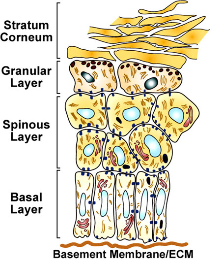 | 452 × 536 (78 KB) | S8600021 (talk | contribs) | ==Epidermal Differentiation== The program of epidermal differentiation is shown in this schematic, illustrating the basement membrane at the base, the proliferative basal layer, and the three differentiation stages: spinous layer, granular layer, and out |
You cannot overwrite this file.
File usage
The following 2 pages use this file: