Template:Corpus Luteum Vignette: Difference between revisions
From Embryology
(Created page with "{| |+ '''Historic Embryology''' |-bgcolor="FAF5FF" | left|100px|link=Embryology History - Reinier de Graaf Em...") |
mNo edit summary |
||
| Line 1: | Line 1: | ||
{| | {| | ||
| | |-bgcolor="DDCEF2" | ||
| [[Historic Embryology Vignette|'''Historic Embryology''']] | |||
|-bgcolor="FAF5FF" | |-bgcolor="FAF5FF" | ||
| [[File:Regnier de Graaf.jpg|left|100px|link=Embryology History - Reinier de Graaf]] | | [[File:Regnier de Graaf.jpg|left|100px|link=Embryology History - Reinier de Graaf]] | ||
Latest revision as of 17:35, 6 February 2020
| Historic Embryology |
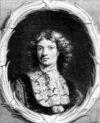 Regnier de Graaf (1641 – 1673) first observed histologically the corpus luteum in the ovary of a cow by its defining yellow structure. The yellow colour is caused by the accumulation of steroidal hormones.
|

