File:Radford1908 fig02.jpg: Difference between revisions
From Embryology
mNo edit summary |
|||
| Line 1: | Line 1: | ||
==Fig. 2. Transverse section of embryo of 10 mm total length== | ==Fig. 2. Transverse section of embryo of 10 mm total length== | ||
Showing | Showing mesenteric artery with surrounding coat of lymphoid cells and the first indication of the {{spleen}} anlage. High power. | ||
BC, blood-corpuscles; CH, notochord ; I, intestine ; IE’, | BC, blood-corpuscles; CH, notochord ; I, intestine ; IE’, strayed endodermal cells; MA, mesenteric artery; MY, myot--me; PLY, primitive lymphoid cells; SPA, spleen anlage. | ||
Revision as of 12:49, 19 July 2019
Fig. 2. Transverse section of embryo of 10 mm total length
Showing mesenteric artery with surrounding coat of lymphoid cells and the first indication of the spleen anlage. High power.
BC, blood-corpuscles; CH, notochord ; I, intestine ; IE’, strayed endodermal cells; MA, mesenteric artery; MY, myot--me; PLY, primitive lymphoid cells; SPA, spleen anlage.
Reference
Radford M. Development of the spleen. (1908) J Anat Physiol. 42: 288-301.
Cite this page: Hill, M.A. (2024, June 7) Embryology Radford1908 fig02.jpg. Retrieved from https://embryology.med.unsw.edu.au/embryology/index.php/File:Radford1908_fig02.jpg
- © Dr Mark Hill 2024, UNSW Embryology ISBN: 978 0 7334 2609 4 - UNSW CRICOS Provider Code No. 00098G
File history
Click on a date/time to view the file as it appeared at that time.
| Date/Time | Thumbnail | Dimensions | User | Comment | |
|---|---|---|---|---|---|
| current | 12:50, 19 July 2019 | 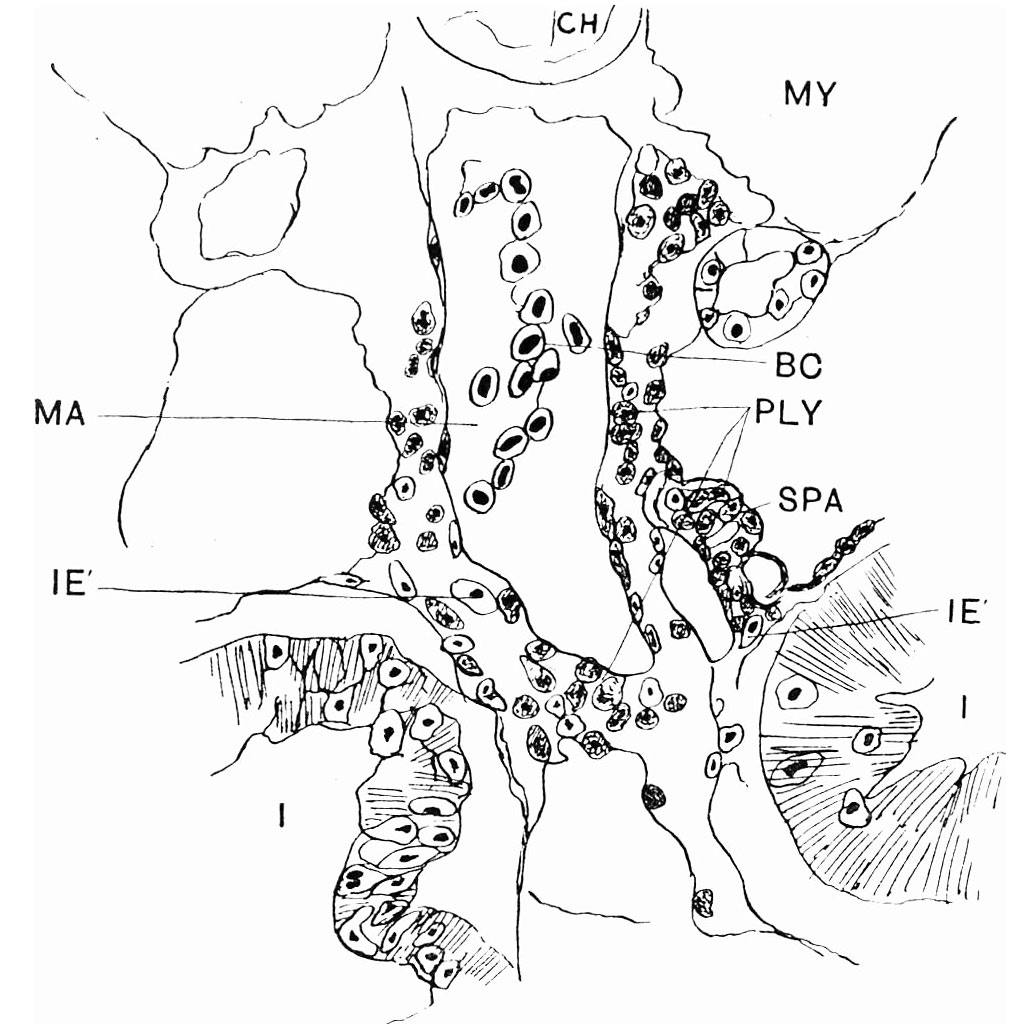 | 1,031 × 1,024 (168 KB) | Z8600021 (talk | contribs) | |
| 12:47, 19 July 2019 | 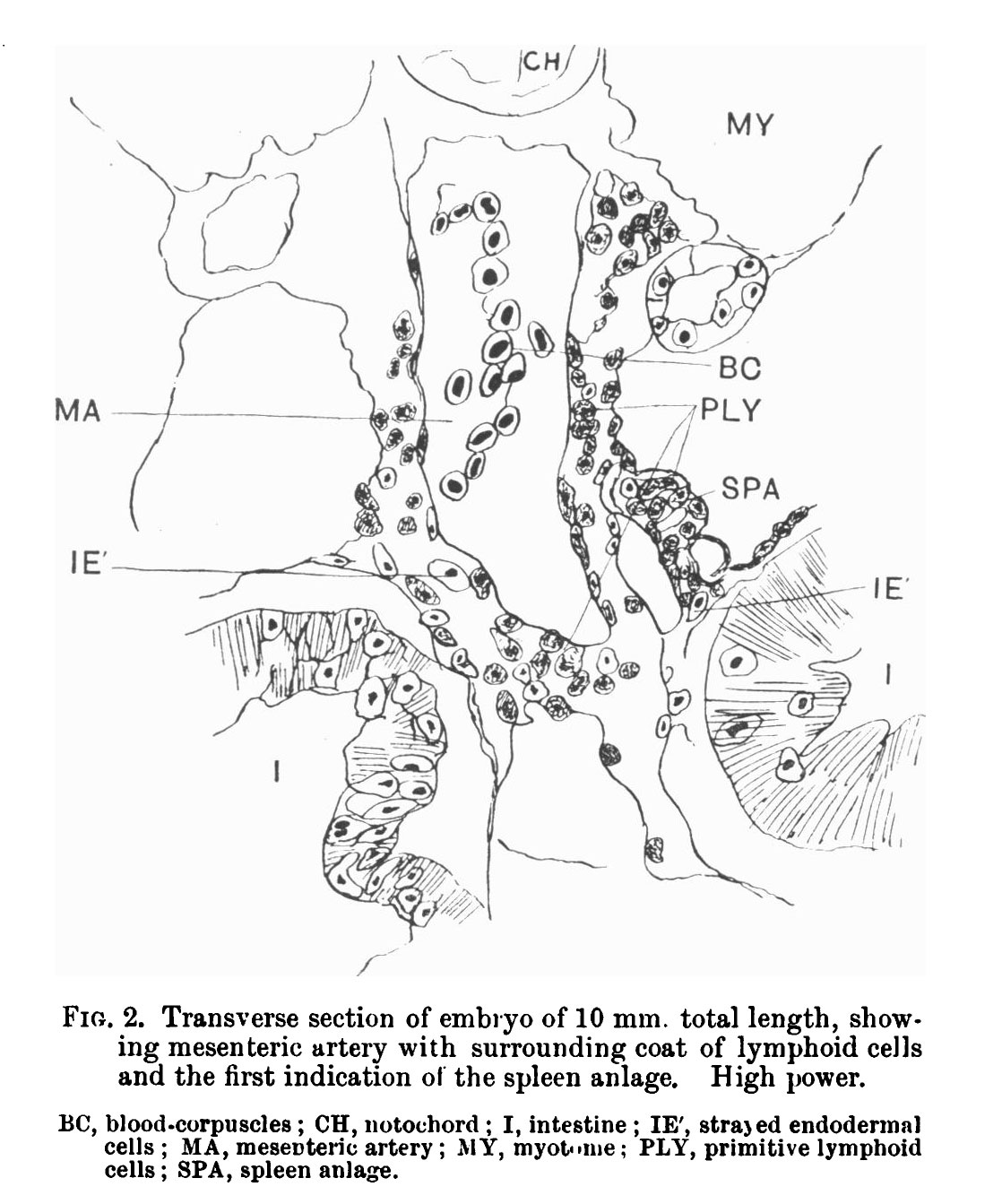 | 1,118 × 1,343 (210 KB) | Z8600021 (talk | contribs) |
You cannot overwrite this file.
File usage
The following page uses this file: