|
|
| Line 30: |
Line 30: |
|
| |
|
| ===Reference=== | | ===Reference=== |
| <pubmed>19644499</pubmed>| [http://www.ncbi.nlm.nih.gov/pmc/articles/PMC2785037 PMC2785037] | [http://www.nature.com/nri/journal/v9/n9/abs/nri2588.html Nat Rev Immunol.]
| | {{#pmid:19644499}} |
|
| |
|
| | ====Copyright==== |
| [http://www.microbiol.unimelb.edu.au/research/immunology/s_mueller.html Mueller] | | [http://www.microbiol.unimelb.edu.au/research/immunology/s_mueller.html Mueller] |
|
| |
|
| [[File_talk:Spleen_structure_01.jpg|Permissions]] | | [[File_talk:Spleen_structure_01.jpg|Permissions]] |
|
| |
|
| | | {{Footer}} |
| [[Category:Spleen]] | | [[Category:Spleen]] |
Latest revision as of 10:03, 19 July 2019
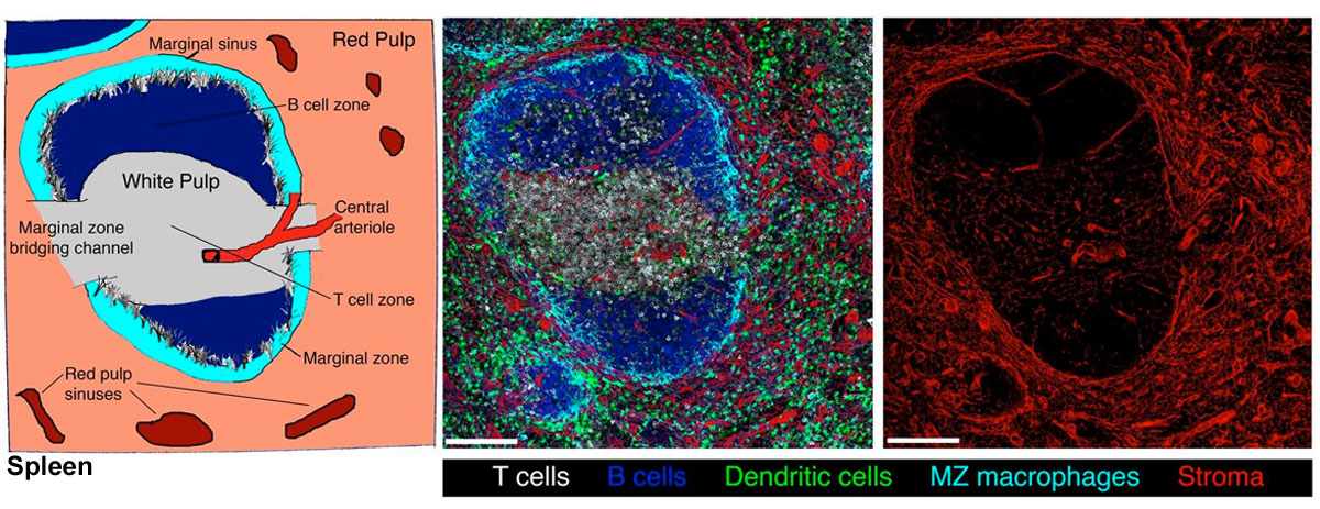
Spleen Structure and Cell Distribution
Schematic representation of the organization of the spleen (left panel).
- The white pulp consists of T cell (lymphocyte) zones (also known as the periarteriolar lymphoid sheath (PALS)) containing networks of fibroblastic reticular cells (FRC) surrounding a central arteriole, together with B cell follicles containing a central network of follicular dendritic cells (FDC).
- Marginal zones (MZ) surrounding the white pulp contain marginal reticular cells (MRC), particularly at the edges of the B cell follicles.
- Blood and leukocytes entering the spleen pass through branches of the central arteriole, which end in the marginal sinuses and red pulp.
- In the cords of the red pulp, a dense network of reticular fibroblasts and fibres construct an open blood network, which is marked by its lack of a typical endothelial cell lining.
- Large numbers of macrophages phagocytose dying or damaged red blood cells in the red pulp (not shown).
- Immune cells enter the white pulp at regions where the T cell zones abut the MZ, known as the MZ bridging channels.
|
An image of a section of mouse spleen generated using multicolour immunofluoresence microscopy illustrates the organization of the white pulp, red pulp, and MZ (centre panel).
- The distribution of cells:
- T cells - (white) CD3+
- B cells - (blue) B220+
- macrophages - (cyan) CD169+ MZ
- dendritic cells - (green) (DCs) CD11c+
- stromal cells - (red) ER-TR7+
- The distinct organization of stromal cells in different regions of the spleen is shown by single-colour immunofluoresence staining (right panel).
- Networks of stromal cells and reticular fibres form in the white pulp, including the fibroblastic reticular cells (FRCs) in T cell zones, follicular dendritic cells (FDCs) in B cell follicles (ER-TR7−) and marginal reticular cells (MRCs) in the MZ.
- A dense network of stromal cells and reticular fibres is present in the red pulp.
Scale bars represent 130 μM.
|
Reference
Mueller SN & Germain RN. (2009). Stromal cell contributions to the homeostasis and functionality of the immune system. Nat. Rev. Immunol. , 9, 618-29. PMID: 19644499 DOI.
Copyright
Mueller
Permissions
Cite this page: Hill, M.A. (2024, June 16) Embryology Spleen structure 01.jpg. Retrieved from https://embryology.med.unsw.edu.au/embryology/index.php/File:Spleen_structure_01.jpg
- What Links Here?
- © Dr Mark Hill 2024, UNSW Embryology ISBN: 978 0 7334 2609 4 - UNSW CRICOS Provider Code No. 00098G
Click on a date/time to view the file as it appeared at that time.
| Date/Time | Thumbnail | Dimensions | User | Comment |
|---|
| current | 18:55, 22 February 2012 |  | 1,200 × 463 (211 KB) | Z8600021 (talk | contribs) | |
You cannot overwrite this file.
The following page uses this file: