File:Human cochlea fetal development cartoon.jpg: Difference between revisions
(Human cochlea fetal development cartoon © 2013 Locher et al.; licensee BioMed Central Ltd. This is an open access article distributed under the terms of the Creative Commons Attribution License (http://creativecommons.org/licenses/by/2.0), which per...) |
|||
| (10 intermediate revisions by the same user not shown) | |||
| Line 1: | Line 1: | ||
Human cochlea fetal development cartoon | ==Human Cochlea Fetal Development Cartoon== | ||
Schematic diagram of neurosensory development in the basal turn of the human fetal cochlea by Gestational Week {{GA}} | |||
{| | |||
| width=600px| | |||
'''Gestational Week''' {{GA}} | |||
* '''Week 10''' (W10) - SOX2 identifies the prosensory domain within the SOX9/SOX10+ cochlear duct epithelium. Neurites from the adjoining TUBB3+/PRPH + SGNs do not yet penetrate into the epithelium. | |||
* '''Week 11''' (W11) - Penetration starts prior to hair cell differentiation. | |||
* '''Week 12''' (W12) - The first MYO7A+/SOX9-/SOX10-/SOX2+ (inner) hair cell can be seen, and is contacted by multiple TUBB3+ and PRPH + neurites. Penetrating neurites are also found at the location of the future OHCs. | |||
* '''Week 14''' (W14) - Both the IHCs and OHCs have differentiated, and neurites underneath the OHCs start to run in a spiral direction. At this stage, hair cells still express SOX2. | |||
* '''Week 20''' (W20) - SOX2 is downregulated in all hair cells, as opposed to the other cells in the organ of Corti. PRPH expression distinguishes between type I (PRPH-) and type II (PRPH+) neurites. | |||
| valign=top|'''Abbreviations''' | |||
* SGN - spiral ganglion neuron | |||
* IHC - inner hair cell | |||
* O1 - first row of outer hair cells | |||
* O2 - second row of outer hair cells | |||
* O3 - third row of outer hair cells | |||
* OHC - outer hair cell. | |||
|} | |||
:[[Hearing_-_Inner_Ear_Development|'''Cochlea Links''']]: [[:File:Human fetal cochlea 01.jpg|Image week 8]] | [[:File:Human fetal cochlea 02.jpg|Image week 10]] | [[:File:Human cochlea fetal development cartoon.jpg|Cochlea molecular cartoon]] | [[Hearing_-_Inner_Ear_Development|Inner Ear Development]] | |||
===Reference=== | |||
{{#pmid:24131517}} | |||
====Copyright==== | |||
© 2013 Locher et al.; licensee BioMed Central Ltd. | © 2013 Locher et al.; licensee BioMed Central Ltd. | ||
This is an open access article distributed under the terms of the Creative Commons Attribution License (http://creativecommons.org/licenses/by/2.0), which permits unrestricted use, distribution, and reproduction in any medium, provided the original work is properly cited. | This is an open access article distributed under the terms of the Creative Commons Attribution License (http://creativecommons.org/licenses/by/2.0), which permits unrestricted use, distribution, and reproduction in any medium, provided the original work is properly cited. | ||
1749-8104-8-20-9.jpg | Figure 9. 1749-8104-8-20-9.jpg Locher et al. Neural Development 2013 8:20 doi:10.1186/1749-8104-8-20 Original figure altered in size and labelling. | ||
{{Footer}} | |||
[[Category:Human]] [[Category:Fetal]] [[Category:Hearing]] [[Category:Inner Ear]][[Category:Week 8]][[Category:Week 9]][[Category:Week 10]][[Category:Week 12]][[Category:Week 18]] | |||
Latest revision as of 21:07, 12 May 2019
Human Cochlea Fetal Development Cartoon
Schematic diagram of neurosensory development in the basal turn of the human fetal cochlea by Gestational Week GA
|
Gestational Week GA
|
Abbreviations
|
Reference
Locher H, Frijns JH, van Iperen L, de Groot JC, Huisman MA & Chuva de Sousa Lopes SM. (2013). Neurosensory development and cell fate determination in the human cochlea. Neural Dev , 8, 20. PMID: 24131517 DOI.
Copyright
© 2013 Locher et al.; licensee BioMed Central Ltd. This is an open access article distributed under the terms of the Creative Commons Attribution License (http://creativecommons.org/licenses/by/2.0), which permits unrestricted use, distribution, and reproduction in any medium, provided the original work is properly cited.
Figure 9. 1749-8104-8-20-9.jpg Locher et al. Neural Development 2013 8:20 doi:10.1186/1749-8104-8-20 Original figure altered in size and labelling.
Cite this page: Hill, M.A. (2024, June 26) Embryology Human cochlea fetal development cartoon.jpg. Retrieved from https://embryology.med.unsw.edu.au/embryology/index.php/File:Human_cochlea_fetal_development_cartoon.jpg
- © Dr Mark Hill 2024, UNSW Embryology ISBN: 978 0 7334 2609 4 - UNSW CRICOS Provider Code No. 00098G
File history
Yi efo/eka'e gwa ebo wo le nyangagi wuncin ye kamina wunga tinya nan
| Gwalagizhi | Nyangagi | Dimensions | User | Comment | |
|---|---|---|---|---|---|
| current | 11:41, 29 June 2014 | 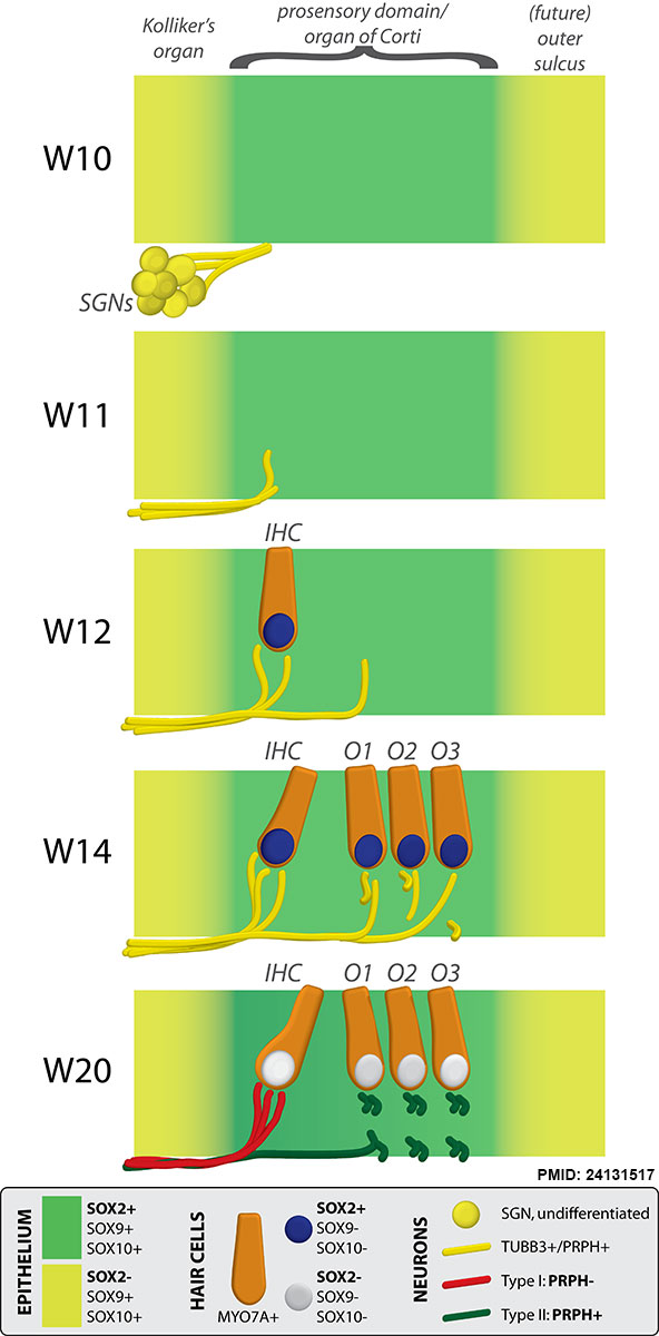 | 592 × 1,200 (96 KB) | Z8600021 (talk | contribs) | Human cochlea fetal development cartoon © 2013 Locher et al.; licensee BioMed Central Ltd. This is an open access article distributed under the terms of the Creative Commons Attribution License (http://creativecommons.org/licenses/by/2.0), which per... |
You cannot overwrite this file.
File usage
The following page uses this file: