File:Lymph node cartoon 02.jpg: Difference between revisions
m (→Copyright) |
|||
| (8 intermediate revisions by the same user not shown) | |||
| Line 2: | Line 2: | ||
Lymphocytes and dendritic cells (DCs) enter lymph nodes by different routes. | Lymphocytes and dendritic cells (DCs) enter lymph nodes by different routes. | ||
{| | |||
| width=500px| | |||
===Lymphocytes=== | ===Lymphocytes=== | ||
* Most lymphocytes migrating to lymph nodes enter from the peripheral blood. | * Most lymphocytes migrating to lymph nodes enter from the peripheral blood. | ||
* All leukocyte types are found in the arteries of lymph nodes. | * All leukocyte types are found in the arteries of lymph nodes. | ||
* Only lymphocytes can interact with and extravasate through high endothelial venules (HEVs) to migrate into the lymph node parenchyma. | * Only lymphocytes can interact with and extravasate through high endothelial venules (HEVs) to migrate into the lymph node parenchyma. | ||
* T and B cells (lymphocytes) subsequently segregate into the T-cell zones and B-cell zones. | * T and B cells (lymphocytes) subsequently segregate into the T-cell zones and B-cell zones. | ||
| | |||
===Legend=== | |||
* '''B''' - B-cell zones | |||
* '''DC''' - dendritic cells | |||
* '''FRC''' - fibroblastic reticular cells | |||
* '''HEV''' - high endothelial venues | |||
|} | |||
===Dendritic cells=== | ===Dendritic cells=== | ||
* DCs | * Dendritic cells (DCs) with small numbers of lymphocytes enter lymph nodes through the afferent lymphatics. | ||
* then accumulate in the vicinity of high endothelial venules. | * then accumulate in the vicinity of high endothelial venules. | ||
* high endothelial venules are surrounded by fibroblastic reticular cells (FRCs) | * high endothelial venules are surrounded by fibroblastic reticular cells (FRCs) | ||
===Fibroblastic reticular cells=== | ===Fibroblastic reticular cells=== | ||
* (FRCs) form channels (FRC conduits) that project from the subcapsular sinus into the T-cell zone. | * (FRCs) form channels (FRC conduits) that project from the subcapsular sinus into the T-cell zone. | ||
* Some chemokines produced extranodally might reach HEVs through the FRC conduit. | * Some chemokines produced extranodally might reach HEVs through the FRC conduit. | ||
See also [[:File:Lymph node - high endothelial venule.jpg|Lymph node - high endothelial venule cartoon]]. | |||
{{Lymph node cartoons}} | {{Lymph node cartoons}} | ||
| Line 31: | Line 43: | ||
===Reference=== | ===Reference=== | ||
{{#pmid:15122201}} | |||
====Copyright==== | |||
Adapted by permission from Macmillan Publishers Ltd: Nat. Rev. Immunol.: 2004, 4(5);360-70, copyright (2004) | Adapted by permission from Macmillan Publishers Ltd: Nat. Rev. Immunol.: 2004, 4(5);360-70, copyright (2004) | ||
| Line 44: | Line 54: | ||
https://s100.copyright.com/CustomerAdmin/PLF.jsp?lID=2012021_1330385293727 | https://s100.copyright.com/CustomerAdmin/PLF.jsp?lID=2012021_1330385293727 | ||
{{Footer}} | |||
[[Category:Blood]][[Category:Immune]][[Category:Lymph Node]] | |||
Latest revision as of 11:24, 12 February 2019
Cell Trafficking into and out of Lymph Nodes
Lymphocytes and dendritic cells (DCs) enter lymph nodes by different routes.
Lymphocytes
|
Legend
|
Dendritic cells
- Dendritic cells (DCs) with small numbers of lymphocytes enter lymph nodes through the afferent lymphatics.
- then accumulate in the vicinity of high endothelial venules.
- high endothelial venules are surrounded by fibroblastic reticular cells (FRCs)
Fibroblastic reticular cells
- (FRCs) form channels (FRC conduits) that project from the subcapsular sinus into the T-cell zone.
- Some chemokines produced extranodally might reach HEVs through the FRC conduit.
See also Lymph node - high endothelial venule cartoon.
- Lymph Node Cartoons: Detailed structure | Cartoon with Histology | Lymphocyte traffic | Simple structure | Simple node anatomy | Wiki node image | Internal structure | Mesenteric lymph node | Histology | Gallery | Lymph Node Development
Reference
Miyasaka M & Tanaka T. (2004). Lymphocyte trafficking across high endothelial venules: dogmas and enigmas. Nat. Rev. Immunol. , 4, 360-70. PMID: 15122201 DOI.
Copyright
Adapted by permission from Macmillan Publishers Ltd: Nat. Rev. Immunol.: 2004, 4(5);360-70, copyright (2004)
Licensee: Mark A Hill License Number: 2857291045727 Publication: Nature Reviews Immunology
Title: Lymphocyte trafficking across high endothelial venules: dogmas and enigmas Type Of Use: post on the internet
https://s100.copyright.com/CustomerAdmin/PLF.jsp?lID=2012021_1330385293727
Cite this page: Hill, M.A. (2024, June 3) Embryology Lymph node cartoon 02.jpg. Retrieved from https://embryology.med.unsw.edu.au/embryology/index.php/File:Lymph_node_cartoon_02.jpg
- © Dr Mark Hill 2024, UNSW Embryology ISBN: 978 0 7334 2609 4 - UNSW CRICOS Provider Code No. 00098G
File history
Click on a date/time to view the file as it appeared at that time.
| Date/Time | Thumbnail | Dimensions | User | Comment | |
|---|---|---|---|---|---|
| current | 09:56, 28 February 2012 | 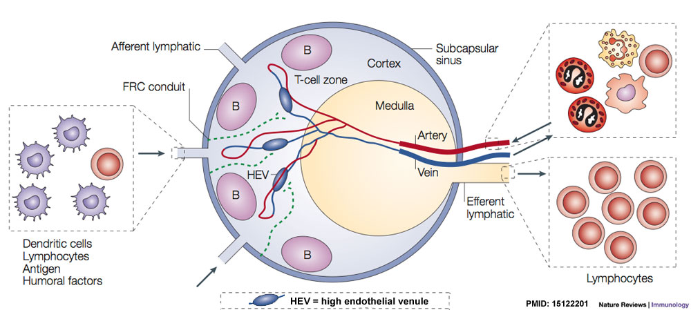 | 1,000 × 449 (97 KB) | Z8600021 (talk | contribs) | |
| 09:41, 28 February 2012 |  | 1,000 × 449 (95 KB) | Z8600021 (talk | contribs) | {{Lymph node cartoons}} |
You cannot overwrite this file.