File:Drg SKETCH.jpg: Difference between revisions
No edit summary |
mNo edit summary |
||
| Line 2: | Line 2: | ||
Note - This image was originally uploaded as part of an undergraduate science student 2018 project and may contain inaccuracies in either description or acknowledgements. Students have been advised in writing concerning the reuse of content and may accidentally have misunderstood the original terms of use. If image reuse on this non-commercial educational site infringes your existing copyright, please contact the site editor for immediate removal. | Note - This image was originally uploaded as part of an undergraduate science student 2018 project and may contain inaccuracies in either description or acknowledgements. Students have been advised in writing concerning the reuse of content and may accidentally have misunderstood the original terms of use. If image reuse on this non-commercial educational site infringes your existing copyright, please contact the site editor for immediate removal. | ||
[[User:Z8600021|Mark Hill]] ([[User talk:Z8600021|talk]]) 09:56, 18 October 2018 (AEDT) Student drawn images in a project are always a welcome addition. There should have been additional descriptive information here as well as the original source upon which your drawing is based. I am also wondering what additional information this figure shows compared to the other figure on the project page - [[:File:Dorsal Root Ganglia Adult.jpg|Dorsal Root Ganglia Adult]]? | |||
Latest revision as of 08:56, 18 October 2018
student sketch showing location and structure of dorsal root ganglion in association to the spinal cord of humans.
Note - This image was originally uploaded as part of an undergraduate science student 2018 project and may contain inaccuracies in either description or acknowledgements. Students have been advised in writing concerning the reuse of content and may accidentally have misunderstood the original terms of use. If image reuse on this non-commercial educational site infringes your existing copyright, please contact the site editor for immediate removal.
Mark Hill (talk) 09:56, 18 October 2018 (AEDT) Student drawn images in a project are always a welcome addition. There should have been additional descriptive information here as well as the original source upon which your drawing is based. I am also wondering what additional information this figure shows compared to the other figure on the project page - Dorsal Root Ganglia Adult?
File history
Yi efo/eka'e gwa ebo wo le nyangagi wuncin ye kamina wunga tinya nan
| Gwalagizhi | Nyangagi | Dimensions | User | Comment | |
|---|---|---|---|---|---|
| current | 17:09, 17 September 2018 | 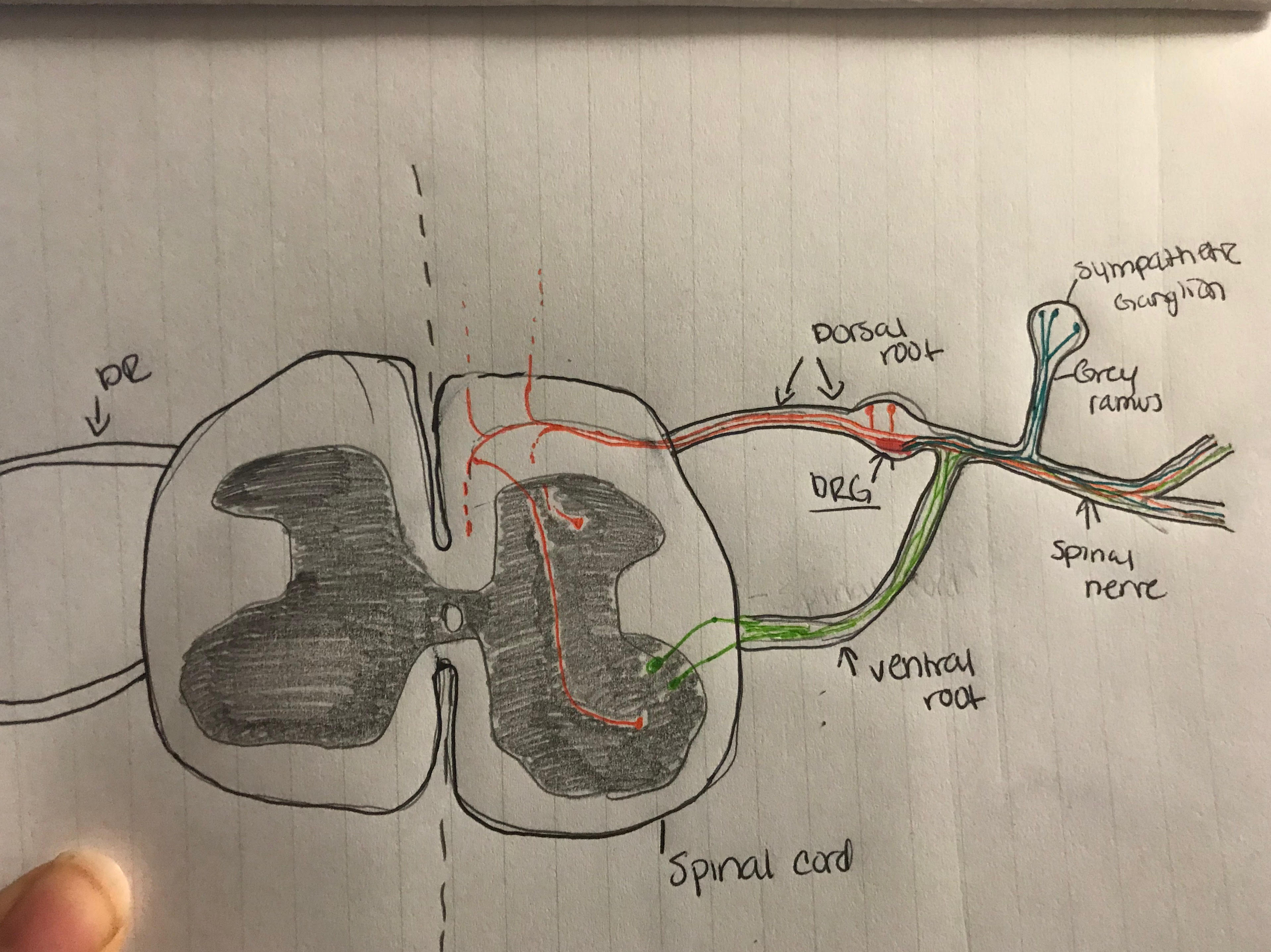 | 3,836 × 2,874 (1.26 MB) | Z5229438 (talk | contribs) | student sketch showing location and structure of dorsal root ganglion in association to the spinal cord of humans. |
You cannot overwrite this file.
File usage
The following 2 pages use this file: