File:Leydig cell PMID13693345 EM02.jpg: Difference between revisions
mNo edit summary |
m (→Reference) |
||
| Line 11: | Line 11: | ||
===Reference=== | ===Reference=== | ||
{{#pmid:13693345}} | |||
{{JCB}} | {{JCB}} | ||
Original figure 3 rotated, relabelled and adjusted in size and contrast. | Original figure 3 rotated, relabelled and adjusted in size and contrast. | ||
Revision as of 14:41, 23 July 2018
Leydig Cell EM
Opossum testicular interstitial cells showing extensive smooth endoplasmic reticulum involved in testosterone synthesis.
A low-power electron micrograph showing several interstitial cells, one of which (above) contains cytoplasm of considerably greater density than the others. The most striking feature of the interstitial cells is the abundant agranular endoplasmic reticulmn (agr), which fills their cytoplasm and is in the form of a network of interconnected tubules. At the periphery of thc cells is an ectoplasmic zone (ect), which is relatively free of endoplasmic reticulum. The cytoplasm also contains mitochondria (m), in which large, homogeneous granules (g) sometimes occur (see Fig. 10). Various extensions of the Golgi zone (G) arc seen around the nucleus. Small, dark bodies (b) of unknown nature are also found in the cytoplasm. The denser cell contains an oblique section through a bundle (bun) of minute tubules, but it is difficult to make out detail because of the great density of the cytoplasm.
The edge of a seminiferous tubule (sere) is seen at lower right, and is flanked by one of the cells (lain) which contribute to the lamina propria of the tubule. X 6,200.
Reference
CHRISTENSEN AK & FAWCETT DW. (1961). The normal fine structure of opossum testicular interstitial cells. J Biophys Biochem Cytol , 9, 653-70. PMID: 13693345
Copyright
Rockefeller University Press - Copyright Policy This article is distributed under the terms of an Attribution–Noncommercial–Share Alike–No Mirror Sites license for the first six months after the publication date (see http://www.jcb.org/misc/terms.shtml). After six months it is available under a Creative Commons License (Attribution–Noncommercial–Share Alike 4.0 Unported license, as described at https://creativecommons.org/licenses/by-nc-sa/4.0/ ). (More? Help:Copyright Tutorial)
Original figure 3 rotated, relabelled and adjusted in size and contrast.
File history
Click on a date/time to view the file as it appeared at that time.
| Date/Time | Thumbnail | Dimensions | User | Comment | |
|---|---|---|---|---|---|
| current | 17:13, 7 August 2014 | 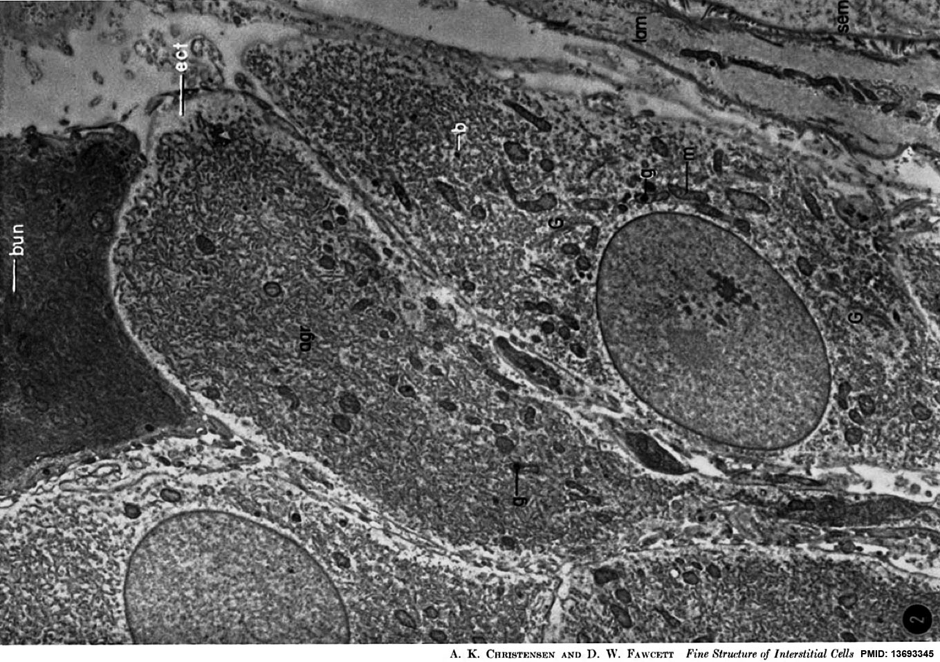 | 1,359 × 957 (341 KB) | Z8600021 (talk | contribs) | J Biophys Biochem Cytol. 1961 Mar;9:653-70. The normal fine structure of opossum testicular interstitial cells. CHRISTENSEN AK, FAWCETT DW. Abstract The interstitial tissue of the opossum testis includes interstitial or Leydig cells, macrophages, and s... |
You cannot overwrite this file.
File usage
The following page uses this file: