File:Shanklin1940 fig04.jpg: Difference between revisions
From Embryology
mNo edit summary |
(Z8600021 uploaded a new version of File:Shanklin1940 fig04.jpg) |
(No difference)
| |
Revision as of 22:20, 26 Haziran 2018
Fig. 4. Photomicrograph through the pars nervosa of a 55-month human foetus
Showing the differentiated pituicytes in the part proximal to the pars intermedia and the poorly differentiated pituicytes in the distal portion. Note a single clump of blood-vessels deep in the nervosa. x 175. I
Reference
Shanklin WM. Differentiation of pituicytes in the human foetus. (1940) J Anat. 74(4): 459-63. PMID 17104829
Cite this page: Hill, M.A. (2026, October 30) Embryology Shanklin1940 fig04.jpg. Retrieved from https://embryology.med.unsw.edu.au/embryology/index.php/File:Shanklin1940_fig04.jpg
- © Dr Mark Hill 2026, UNSW Embryology ISBN: 978 0 7334 2609 4 - UNSW CRICOS Provider Code No. 00098G
File history
Yi efo/eka'e gwa ebo wo le nyangagi wuncin ye kamina wunga tinya nan
| Gwalagizhi | Nyangagi | Dimensions | User | Comment | |
|---|---|---|---|---|---|
| current | 22:20, 26 Haziran 2018 |  | 884 × 1,018 (112 KB) | Z8600021 (talk | contribs) | |
| 21:59, 26 Haziran 2018 | 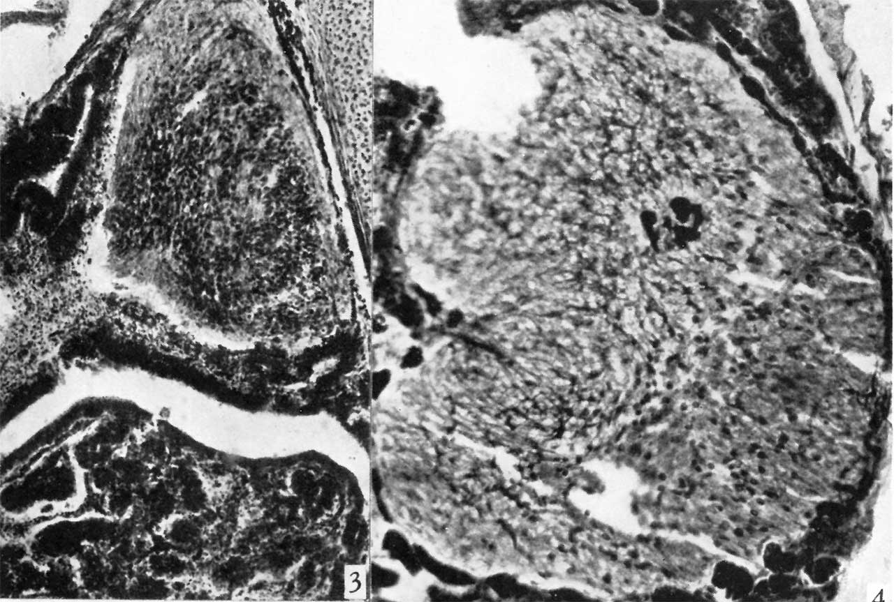 | 1,280 × 863 (162 KB) | Z8600021 (talk | contribs) | ==Plate 1== Fig. 4. Photomicrograph through the pars nervosa of a 55-month human foetus showing the differentiated pituicytes in the part proximal to the pars intermedia and the poorly differentiated pituicytes in the distal portion. Note a single clu... |
You cannot overwrite this file.
File usage
The following page uses this file:


