Category:Cat: Difference between revisions
From Embryology
No edit summary |
mNo edit summary |
||
| Line 2: | Line 2: | ||
Main Page - [[Cat Development|'''Cat Development''']] | |||
{{Animal Links}} | |||
[[Category:Animal Development]] | [[Category:Animal Development]] | ||
Revision as of 10:06, 14 April 2018
Pages in category 'Cat'
The following 50 pages are in this category, out of 50 total.
P
- Paper - A study of pathological cat embryos (1909)
- Paper - Abdominal pregnancy in a cat (1932)
- Paper - Development and homology of the mammalian cerebellar fissures 2
- Paper - Models of the pancreas in embryos of the pig, rabbit, cat, and man (1908)
- Paper - Studies on the fine structure of the mammalian testis 1
- Paper - The development of the mammalian pituitary and its morphological significance (1908)
- Paper - The development of the neuraxis in the domestic cat to the stage of twenty-one somites
- Paper - The developmental significance of the mammalian pharyngeal tonsil - Cat
- Paper - The early development of the cat (1924)
- Paper - The early development of the mammalian sternum
- Paper - The fusion of the cardiac anlages and the formation of the cardiac loop in the cat (1916)
- Paper - The histological appearances of the mammalian pituitary body (1908)
- Paper - The Mammalian Cerebellum part 1 (1895)
- Paper - The morphogenesis of the mammalian ovary (1913)
- Paper - The origin of the heart and blood vessels in felis domestica (1924)
- Paper - The pharyngeal pouches and their derivatives in the mammalia
R
- Template:Ref-Brown1913
- Template:Ref-BurgosFawcett1955
- Template:Ref-Coulter1909
- Template:Ref-Hill1924
- Template:Ref-Hunter1932
- Template:Ref-Huntington1911
- Template:Ref-HuntingtonMcClure1906
- Template:Ref-HuntingtonMcClure1907
- Template:Ref-HuntingtonMcClure1910
- Template:Ref-Jordan1909b
- Template:Ref-Kingsbury1913
- Template:Ref-Kingsbury1914
- Template:Ref-Kingsbury1914a
- Template:Ref-Kingsbury1932
- Template:Ref-Schulte1914
- Template:Ref-Schulte1916
- Template:Ref-SchulteTilney1915
- Template:Ref-Stroud1895
- Template:Ref-Thyng1908
- Template:Ref-Watson1924
- Template:Ref-Wislocki1920
T
Media in category 'Cat'
The following 70 files are in this category, out of 70 total.
 Anson-1934 fig08-21.jpg 1,000 × 1,435; 178 KB
 Anson-1934 fig08.jpg 496 × 560; 36 KB
 Anson-1934 fig09.jpg 334 × 491; 17 KB
 Anson-1934 fig10.jpg 310 × 566; 21 KB
 Anson-1934 fig11.jpg 368 × 411; 16 KB
 Anson-1934 fig12.jpg 231 × 571; 19 KB
 Anson-1934 fig13.jpg 276 × 379; 15 KB
 Anson-1934 fig14.jpg 257 × 280; 11 KB
 Anson-1934 fig15.jpg 217 × 198; 7 KB
 Anson-1934 fig16.jpg 296 × 263; 10 KB
 Bailey296 297.jpg 513 × 726; 72 KB
 Bailey331.jpg 890 × 782; 118 KB
 Boyden1931 fig03.jpg 541 × 878; 56 KB
 BurgosFawcett1955 fig11.jpg 1,453 × 2,015; 528 KB
 BurgosFawcett1955 fig13.jpg 1,460 × 2,049; 501 KB
 BurgosFawcett1955 fig14.jpg 1,456 × 1,965; 381 KB
 BurgosFawcett1955 text-fig01.jpg 1,280 × 1,137; 143 KB
 Cameron1916 fig01.jpg 586 × 1,000; 108 KB
 Cardiac muscle EM01.jpg 1,072 × 735; 231 KB
 Cardiac muscle EM02.jpg 1,072 × 735; 224 KB
 Cardiac muscle EM03.jpg 849 × 615; 135 KB
 Cardiac muscle EM04.jpg 1,000 × 680; 191 KB
 Cardiac Muscle EM05.jpg 992 × 733; 158 KB
 Cat 6 toes.jpg 420 × 280; 13 KB
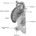
 Cat embryo ovary.jpg 505 × 492; 47 KB
 Cat inner ear MicroCT.jpg 1,159 × 1,300; 266 KB
 Cat oocyte calcium concentration.jpg 600 × 486; 80 KB
 Cat spermatozoa bound to oocyte zona pellucida.jpg 1,000 × 917; 161 KB
 Fox1908 fig62.jpg 725 × 374; 79 KB
 Fox1908 fig63.jpg 1,000 × 787; 98 KB
 Fox1908 fig64.jpg 801 × 791; 82 KB
 Gray0903.jpg 600 × 389; 42 KB
 Gray0939.jpg 681 × 371; 90 KB
 Gray1112.jpg 550 × 548; 51 KB
 Hill1924 plate24.jpg 1,495 × 1,905; 441 KB
 Hill1924 plate25.jpg 1,495 × 1,905; 365 KB
 Hill1924 plate26.jpg 1,495 × 1,905; 483 KB
 Hill1924 plate27-1.jpg 1,495 × 1,905; 278 KB
 Hill1924 plate27-2.jpg 1,495 × 1,905; 257 KB
 Hill1924 plate28-1.jpg 1,495 × 1,905; 248 KB
 Hill1924 plate28-2.jpg 1,495 × 1,905; 198 KB
 Hill1924 plate29.jpg 1,495 × 1,905; 215 KB
 Keibel Mall 2 366.jpg 1,000 × 1,195; 222 KB
 Keith1921 fig091.jpg 919 × 812; 216 KB
 Kingsbury1932 fig1.jpg 1,000 × 1,047; 113 KB
 Kingsbury1932 fig2.jpg 700 × 466; 72 KB
 Kingsbury1932 plate01.jpg 1,280 × 1,977; 356 KB
 Kingsbury1932 plate02.jpg 1,280 × 1,981; 447 KB
 Kingsbury1932 plate03.jpg 1,280 × 1,958; 404 KB
 Kingsbury1932 plate04.jpg 1,280 × 1,960; 426 KB
 Mall Meyer1921 fig210.jpg 240 × 379; 17 KB
 Mall Meyer1921 fig211.jpg 212 × 470; 18 KB
 Meyer1914 fig04-05.jpg 1,200 × 639; 124 KB
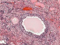 Ovary- atretic follicle 01.jpg 793 × 595; 225 KB
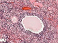 Ovary- atretic follicle 02.jpg 600 × 450; 139 KB
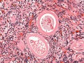 Ovary- atretic follicle 03.jpg 790 × 593; 202 KB
 Ovary- atretic follicle 04.jpg 600 × 450; 128 KB
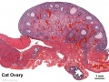 Ovary- histology overview.jpg 861 × 646; 160 KB
 Ovary10x.jpg 480 × 400; 53 KB
 Ovary5x.gif 480 × 400; 162 KB
 Spleen histology 09.jpg 1,280 × 1,024; 692 KB
 Spleen histology 10.jpg 1,280 × 1,024; 444 KB
 Spleen histology 11.jpg 1,280 × 1,024; 410 KB
 Spleen histology 12.jpg 600 × 450; 93 KB
 Spleen histology 13.jpg 600 × 450; 80 KB
 Stomach histology 004.jpg 1,280 × 1,024; 435 KB
 Stomach histology 005.jpg 1,280 × 1,024; 432 KB
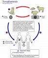 Toxoplasmosis lifecycle.jpg 600 × 729; 75 KB
 Williams1908-fig19.jpg 981 × 1,500; 104 KB
 Wislocki1920 plate 4.jpg 935 × 1,000; 199 KB


