File:Skull CT abnormal 02.jpg: Difference between revisions
No edit summary |
mNo edit summary |
||
| (2 intermediate revisions by the same user not shown) | |||
| Line 2: | Line 2: | ||
* There is complete fusion of the coronal suture (white arrows) with a prominent frontal bone and flattened occiput. | * There is complete fusion of the coronal suture (white arrows) with a prominent frontal bone and flattened occiput. | ||
* Coronal reconstruction (D) demonstrates prominent bilateral elliptical orbits, known as the "harlequin eye" deformity. | * Coronal reconstruction (D) demonstrates prominent bilateral elliptical orbits, known as the "harlequin eye" deformity. | ||
* | * arrowheads in E show the early partial fusion of the right coronal suture. | ||
{{Template:Skull_CT_links}} | |||
===Legend=== | ===Legend=== | ||
| Line 12: | Line 14: | ||
===Reference=== | ===Reference=== | ||
{{#pmid:21431034}} | |||
====Copyright==== | |||
This is an open-access article distributed under the terms of the Creative Commons Attribution License, which permits unrestricted use, distribution, and reproduction in any medium, provided the original work is properly cited. | This is an open-access article distributed under the terms of the Creative Commons Attribution License, which permits unrestricted use, distribution, and reproduction in any medium, provided the original work is properly cited. | ||
| Line 26: | Line 29: | ||
http://www.ijri.org/viewimage.asp?img=IndianJRadiolImaging_2011_21_1_49_76055_f4.jpg | http://www.ijri.org/viewimage.asp?img=IndianJRadiolImaging_2011_21_1_49_76055_f4.jpg | ||
{{Footer}} | |||
[[Category:Human]] [[Category:Skull]] [[Category:Abnormal Development]] [[Category:Computed Tomography]] | [[Category:Human]] [[Category:Skull]] [[Category:Abnormal Development]] [[Category:Computed Tomography]] | ||
Latest revision as of 23:09, 21 March 2018
Skull Coronal Synostosis
- There is complete fusion of the coronal suture (white arrows) with a prominent frontal bone and flattened occiput.
- Coronal reconstruction (D) demonstrates prominent bilateral elliptical orbits, known as the "harlequin eye" deformity.
- arrowheads in E show the early partial fusion of the right coronal suture.
- Skull CT Images: Normal overview | Normal vertex and lateral | Normal endocranial and vertex | Normal Vertex - Fontanels | Dolichocephaly and Scaphocephaly | Coronal Synostosis | Anterior Plagiocephaly | Turricephaly | Posterior Plagiocephaly | Deformational Plagiocepahly | Trigonocephaly | Oxycephaly | Computed Tomography
Legend
- A-C - Bilateral coronal synostosis 3DCT volume rendered images.
- D - Bilateral coronal synostosis, coronal CT scan.
- E - unilateral partial coronal synostosis 3DCT volume rendered images
Reference
Khanna PC, Thapa MM, Iyer RS & Prasad SS. (2011). Pictorial essay: The many faces of craniosynostosis. Indian J Radiol Imaging , 21, 49-56. PMID: 21431034 DOI.
Copyright
This is an open-access article distributed under the terms of the Creative Commons Attribution License, which permits unrestricted use, distribution, and reproduction in any medium, provided the original work is properly cited.
Paritosh C Khanna © 2007 - 2012 Indian Journal of Radiology and Imaging
Attribution-NonCommercial-ShareAlike 3.0 Unported (CC BY-NC-SA 3.0)
Original file name: Figure 3 (A-E) Original figure has been modified, resized and relabelled.
http://www.ijri.org/viewimage.asp?img=IndianJRadiolImaging_2011_21_1_49_76055_f4.jpg
Cite this page: Hill, M.A. (2024, June 26) Embryology Skull CT abnormal 02.jpg. Retrieved from https://embryology.med.unsw.edu.au/embryology/index.php/File:Skull_CT_abnormal_02.jpg
- © Dr Mark Hill 2024, UNSW Embryology ISBN: 978 0 7334 2609 4 - UNSW CRICOS Provider Code No. 00098G
File history
Yi efo/eka'e gwa ebo wo le nyangagi wuncin ye kamina wunga tinya nan
| Gwalagizhi | Nyangagi | Dimensions | User | Comment | |
|---|---|---|---|---|---|
| current | 11:10, 17 March 2012 | 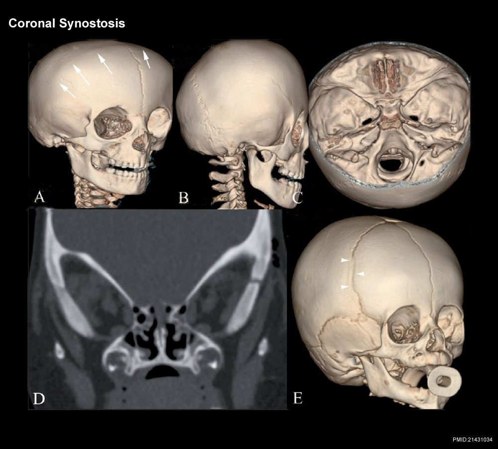 | 1,000 × 900 (119 KB) | Z8600021 (talk | contribs) |
You cannot overwrite this file.
File usage
The following 4 pages use this file: