File:Simkins1928 plate07.jpg: Difference between revisions
({{Simkins1928 figures}}) |
(Z8600021 uploaded a new version of File:Simkins1928 plate07.jpg) |
||
| (One intermediate revision by the same user not shown) | |||
| Line 1: | Line 1: | ||
27 Detail of the large and small cells in the testis of a 54-mm. embryo. X 1500. | |||
28 Secondary follicle from an ovary at term. X 750. A nucleus could not be found in the ovum. | |||
29 Large and small cells in detail from the ovary of a 90-mm. fetus. X 750. | |||
30 Grenitaloid cell in detail from the ovary of a 51-mm. embryo. )< 1500. | |||
31 First indication of a cytoplasmic membrane (vitelline membrane) enclosing the ovum. From an ovary at term. X 350. | |||
32 Mature ovum in the follicle of a full—term ovary: the walls of the follicle have not been shown. X 350. /m.vt., vitelline membrane. | |||
33 Detail of tubule from a 54-mm. embryo. X 1500. cl.so., interstitial cell. | |||
{{Simkins1928 figures}} | {{Simkins1928 figures}} | ||
Latest revision as of 16:54, 31 January 2018
27 Detail of the large and small cells in the testis of a 54-mm. embryo. X 1500.
28 Secondary follicle from an ovary at term. X 750. A nucleus could not be found in the ovum.
29 Large and small cells in detail from the ovary of a 90-mm. fetus. X 750.
30 Grenitaloid cell in detail from the ovary of a 51-mm. embryo. )< 1500.
31 First indication of a cytoplasmic membrane (vitelline membrane) enclosing the ovum. From an ovary at term. X 350.
32 Mature ovum in the follicle of a full—term ovary: the walls of the follicle have not been shown. X 350. /m.vt., vitelline membrane.
33 Detail of tubule from a 54-mm. embryo. X 1500. cl.so., interstitial cell.
| Historic Disclaimer - information about historic embryology pages |
|---|
 |
- Simkins 1928: plate 1 | plate 2 | plate 3 | plate 4 | plate 5 | plate 6 | plate 7 | plate 8 | plate 9 | plate 10
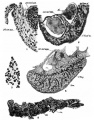
plate 1
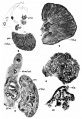
plate 2
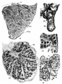
plate 3
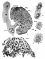
plate 4
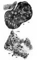
plate 5
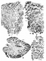
plate 6
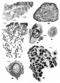
plate 7
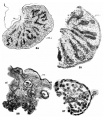
plate 8
plate 9
plate 10


Reference
Simkins CS. Origin of the sex cells in man. (1928) Amer. J Anat. 41: 248-272.
Cite this page: Hill, M.A. (2024, June 27) Embryology Simkins1928 plate07.jpg. Retrieved from https://embryology.med.unsw.edu.au/embryology/index.php/File:Simkins1928_plate07.jpg
- © Dr Mark Hill 2024, UNSW Embryology ISBN: 978 0 7334 2609 4 - UNSW CRICOS Provider Code No. 00098G
File history
Yi efo/eka'e gwa ebo wo le nyangagi wuncin ye kamina wunga tinya nan
| Gwalagizhi | Nyangagi | Dimensions | User | Comment | |
|---|---|---|---|---|---|
| current | 16:54, 31 January 2018 | 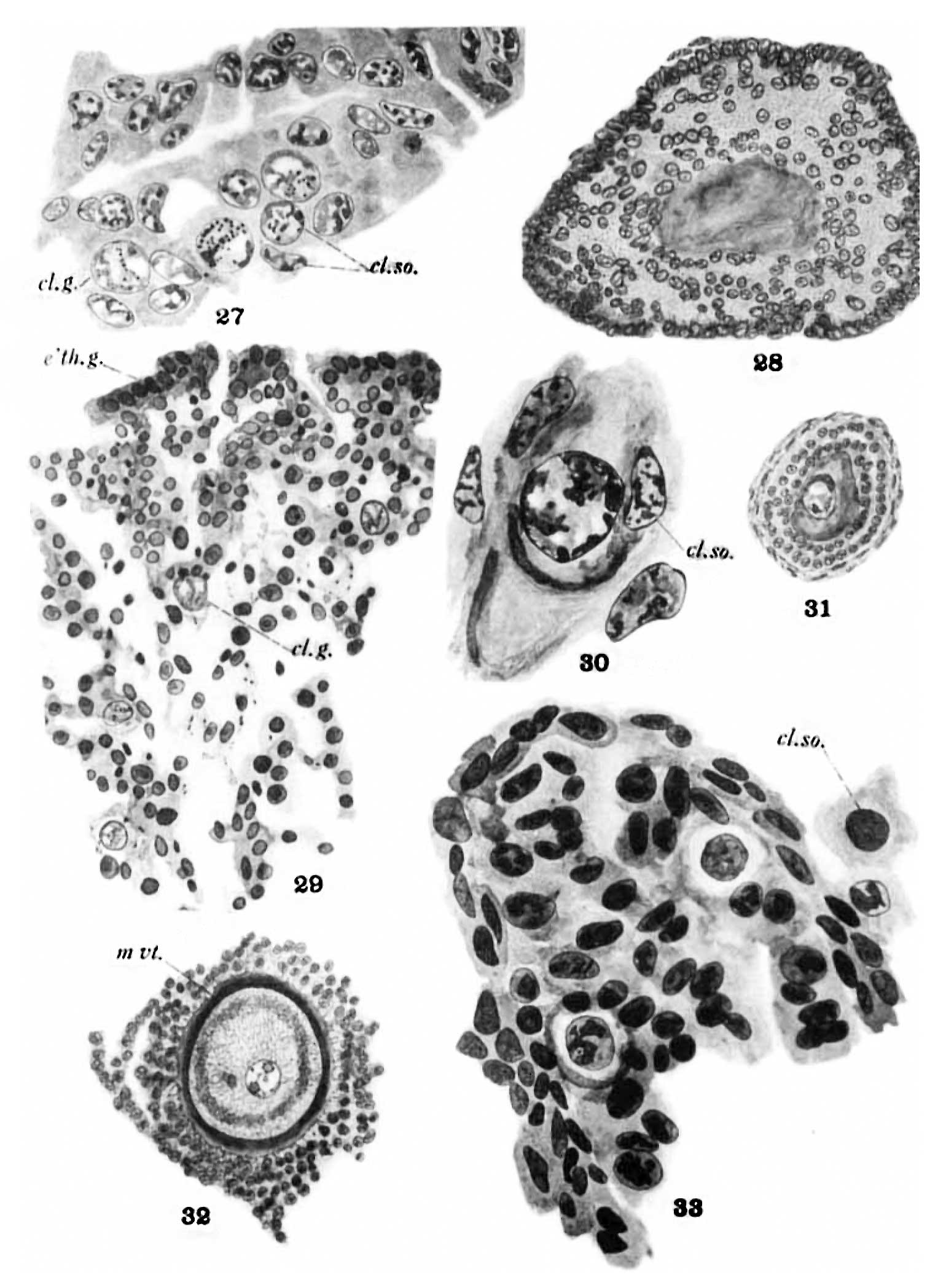 | 1,540 × 2,096 (239 KB) | Z8600021 (talk | contribs) | |
| 16:22, 31 January 2018 | 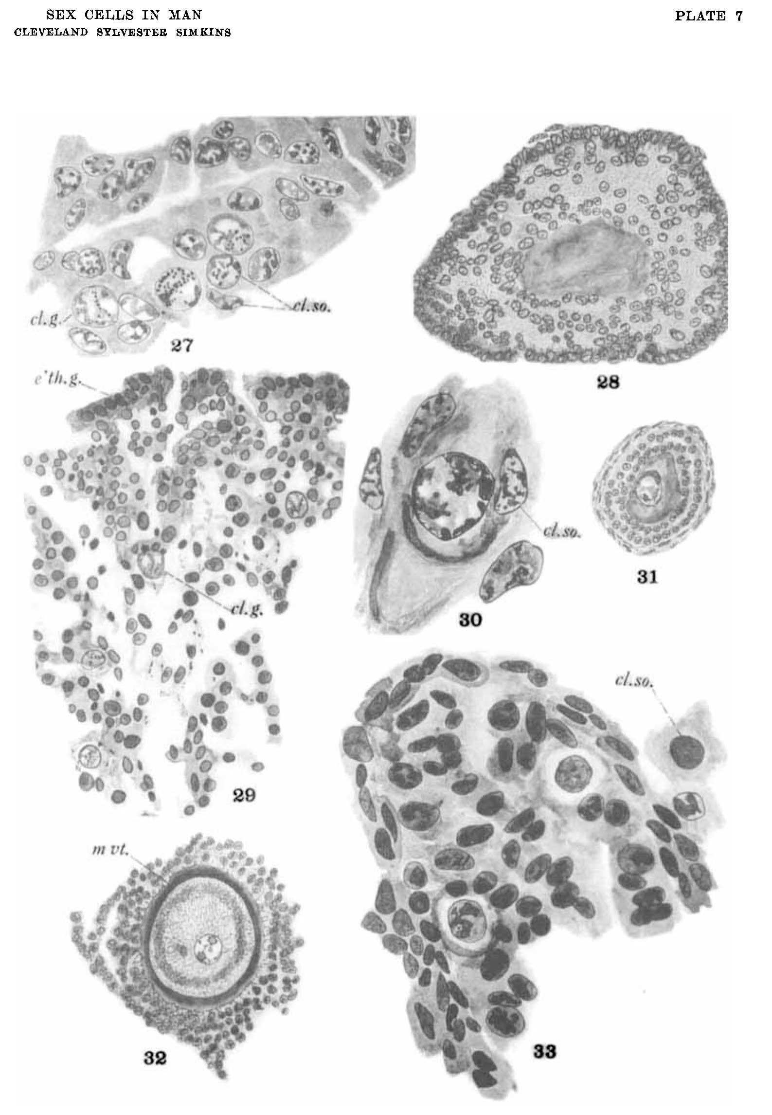 | 1,587 × 2,291 (229 KB) | Z8600021 (talk | contribs) | {{Simkins1928 figures}} |
You cannot overwrite this file.
File usage
The following 12 pages use this file:
- Paper - Origin of the sex cells in man
- File:Simkins1928 plate01.jpg
- File:Simkins1928 plate02.jpg
- File:Simkins1928 plate03.jpg
- File:Simkins1928 plate04.jpg
- File:Simkins1928 plate05.jpg
- File:Simkins1928 plate06.jpg
- File:Simkins1928 plate07.jpg
- File:Simkins1928 plate08.jpg
- File:Simkins1928 plate09.jpg
- File:Simkins1928 plate10.jpg
- Template:Simkins1928 figures