File:Shaw1932 fig04.jpg: Difference between revisions
No edit summary |
mNo edit summary |
||
| Line 1: | Line 1: | ||
==Fig. 4.== | |||
A, cerebral vesicle; B, heart; 0, coelom; D, intestine; E, primitive kidney; F, limb bud; G, dorsal aorta; H, neural canal; C, chorionic villi ; K, amnion; L, umbilical vessel ; M, mesone hric duct; N, neural canal of the embryo , 0, liver. The smaller embryo has no head, heart, or liver. The area of fusion is in the region of the primit ve kidney and umbilicus. The amnion is single. | |||
===Reference=== | |||
{{Ref-Shaw1932}} | |||
{{Footer}} | |||
Latest revision as of 18:09, 15 November 2017
Fig. 4.
A, cerebral vesicle; B, heart; 0, coelom; D, intestine; E, primitive kidney; F, limb bud; G, dorsal aorta; H, neural canal; C, chorionic villi ; K, amnion; L, umbilical vessel ; M, mesone hric duct; N, neural canal of the embryo , 0, liver. The smaller embryo has no head, heart, or liver. The area of fusion is in the region of the primit ve kidney and umbilicus. The amnion is single.
Reference
Shaw W. Observations on two specimens of early human ova. (1932) Brit. Med.J., 1: 411-415.
Cite this page: Hill, M.A. (2026, October 31) Embryology Shaw1932 fig04.jpg. Retrieved from https://embryology.med.unsw.edu.au/embryology/index.php/File:Shaw1932_fig04.jpg
- © Dr Mark Hill 2026, UNSW Embryology ISBN: 978 0 7334 2609 4 - UNSW CRICOS Provider Code No. 00098G
File history
Yi efo/eka'e gwa ebo wo le nyangagi wuncin ye kamina wunga tinya nan
| Gwalagizhi | Nyangagi | Dimensions | User | Comment | |
|---|---|---|---|---|---|
| current | 18:06, 15 November 2017 | 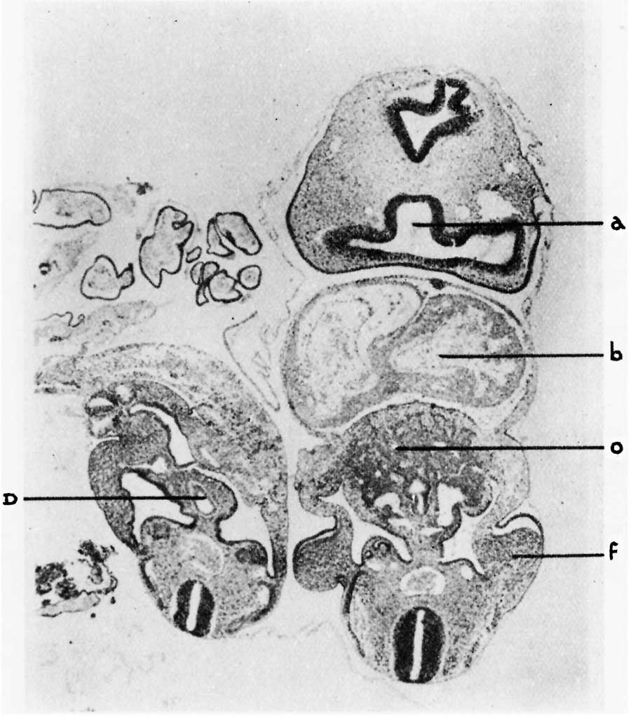 | 900 × 1,021 (100 KB) | Z8600021 (talk | contribs) |
You cannot overwrite this file.
File usage
The following page uses this file:


