File:Bast1933 fig33.jpg: Difference between revisions
mNo edit summary |
mNo edit summary |
||
| (One intermediate revision by the same user not shown) | |||
| Line 4: | Line 4: | ||
Fig. 33. Fetus 86; size, 50 mm. crown-rump length; age, about 10 weeks. Anterior view of a model of the otic labyrinth showing the development of the periotic tissue and spaces and the fissula ante fenestram. | Fig. 33. Fetus 86; size, 50 mm. crown-rump length; age, about 10 weeks. Anterior view of a model of the otic labyrinth showing the development of the periotic tissue and spaces and the fissula ante fenestram. | ||
In figures 33 and 34, vesti. indicates vestibule; f.a.f., fissula ante fenestram; S c. 'vesti., scala vestibuli; Sc. tym., scala tympani; imp. Sta.p., impression of stapes; Sac. end., endolyrnphatic duct; Sm:., sacculus; Ut1'z'c., utriculus; Duct. coch-., cochlear duct. | In figures [[:File:Bast1933 fig33.jpg|33]] and [[:File:Bast1933 fig34.jpg|34]], vesti. indicates vestibule; f.a.f., fissula ante fenestram; S c. 'vesti., scala vestibuli; Sc. tym., scala tympani; imp. Sta.p., impression of stapes; Sac. end., endolyrnphatic duct; Sm:., sacculus; Ut1'z'c., utriculus; Duct. coch-., cochlear duct. | ||
{{Online Editor}} - CRL 50 mm about [[Week 10]] development. | |||
===Reference=== | ===Reference=== | ||
Latest revision as of 12:18, 24 October 2017
Figs. 33 Human Fetus CRL 50 mm
Fig. 33. Fetus 86; size, 50 mm. crown-rump length; age, about 10 weeks. Anterior view of a model of the otic labyrinth showing the development of the periotic tissue and spaces and the fissula ante fenestram.
In figures 33 and 34, vesti. indicates vestibule; f.a.f., fissula ante fenestram; S c. 'vesti., scala vestibuli; Sc. tym., scala tympani; imp. Sta.p., impression of stapes; Sac. end., endolyrnphatic duct; Sm:., sacculus; Ut1'z'c., utriculus; Duct. coch-., cochlear duct.
Online Editor - CRL 50 mm about Week 10 development.
Reference
Bast TH. Development of the otic capsule II. The origin, development and significance of the fissula ante fenestram and its relation to otosclerotic foci. (1933) Arch. Otolaryng. 18(1):
Cite this page: Hill, M.A. (2026, October 31) Embryology Bast1933 fig33.jpg. Retrieved from https://embryology.med.unsw.edu.au/embryology/index.php/File:Bast1933_fig33.jpg
- © Dr Mark Hill 2026, UNSW Embryology ISBN: 978 0 7334 2609 4 - UNSW CRICOS Provider Code No. 00098G
File history
Yi efo/eka'e gwa ebo wo le nyangagi wuncin ye kamina wunga tinya nan
| Gwalagizhi | Nyangagi | Dimensions | User | Comment | |
|---|---|---|---|---|---|
| current | 17:04, 22 October 2017 | 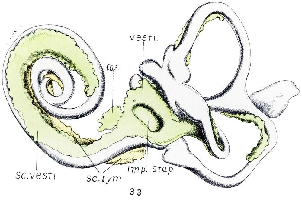 | 1,000 × 673 (73 KB) | Z8600021 (talk | contribs) |
You cannot overwrite this file.
File usage
The following page uses this file:


