File:Anson1948 fig12.jpg: Difference between revisions
No edit summary |
mNo edit summary |
||
| (2 intermediate revisions by the same user not shown) | |||
| Line 1: | Line 1: | ||
==Fig. 12. Drawings of a reconstruction of the stapes in the 180 mm Fetus== | |||
Drawings of a reconstruction of the stapes in the 180 mm. (21 week) fetus (Wisconsin series 45 B); X 7: (a) the entire stapes, in superolateral view; (1)) segment 1 (see inset), viewed from below (that is, here inverted) ; (c) segments 2, 3 and 4 in superomedial view; (d) segment 3, from below (reconstruction inverted), with segment 4 removed; inset, reconstruction with segments lettered and with arrows recording the direction of View in the correspondingly lettered figures. | |||
The drawings show the extent to which periosteal bone has been removed on the obturator surface of the base (a and b), crura and neck (c and d) and the degree to which the stapes throughout has been hollowed (I) to d). Cartilage now remains only on the articular surface of the head and on the vestibular surface and fenestral margin of the base of the ossicle (b and c). The addition of endochondral bone has converted the base into a thinned bilaminar plate ((2 and c). Such bone appears in limited amount as spicules in the area where the crura join the neck of the stapes (d). | |||
===Reference=== | |||
{{Ref-Anson1948}} | |||
{{Footer}} | |||
[[Category:Middle Ear]][[Category:Historic Embryology]][[Category:1940's]] | |||
Latest revision as of 20:52, 16 October 2017
Fig. 12. Drawings of a reconstruction of the stapes in the 180 mm Fetus
Drawings of a reconstruction of the stapes in the 180 mm. (21 week) fetus (Wisconsin series 45 B); X 7: (a) the entire stapes, in superolateral view; (1)) segment 1 (see inset), viewed from below (that is, here inverted) ; (c) segments 2, 3 and 4 in superomedial view; (d) segment 3, from below (reconstruction inverted), with segment 4 removed; inset, reconstruction with segments lettered and with arrows recording the direction of View in the correspondingly lettered figures.
The drawings show the extent to which periosteal bone has been removed on the obturator surface of the base (a and b), crura and neck (c and d) and the degree to which the stapes throughout has been hollowed (I) to d). Cartilage now remains only on the articular surface of the head and on the vestibular surface and fenestral margin of the base of the ossicle (b and c). The addition of endochondral bone has converted the base into a thinned bilaminar plate ((2 and c). Such bone appears in limited amount as spicules in the area where the crura join the neck of the stapes (d).
Reference
Anson BJ. and Cauldwell EW. Stapes, fissula ante fenestram and associated structures in man: V . From the fetus of 160 mm to term. (1948) 48(3): 263-300.
Cite this page: Hill, M.A. (2024, June 27) Embryology Anson1948 fig12.jpg. Retrieved from https://embryology.med.unsw.edu.au/embryology/index.php/File:Anson1948_fig12.jpg
- © Dr Mark Hill 2024, UNSW Embryology ISBN: 978 0 7334 2609 4 - UNSW CRICOS Provider Code No. 00098G
File history
Yi efo/eka'e gwa ebo wo le nyangagi wuncin ye kamina wunga tinya nan
| Gwalagizhi | Nyangagi | Dimensions | User | Comment | |
|---|---|---|---|---|---|
| current | 20:51, 16 October 2017 | 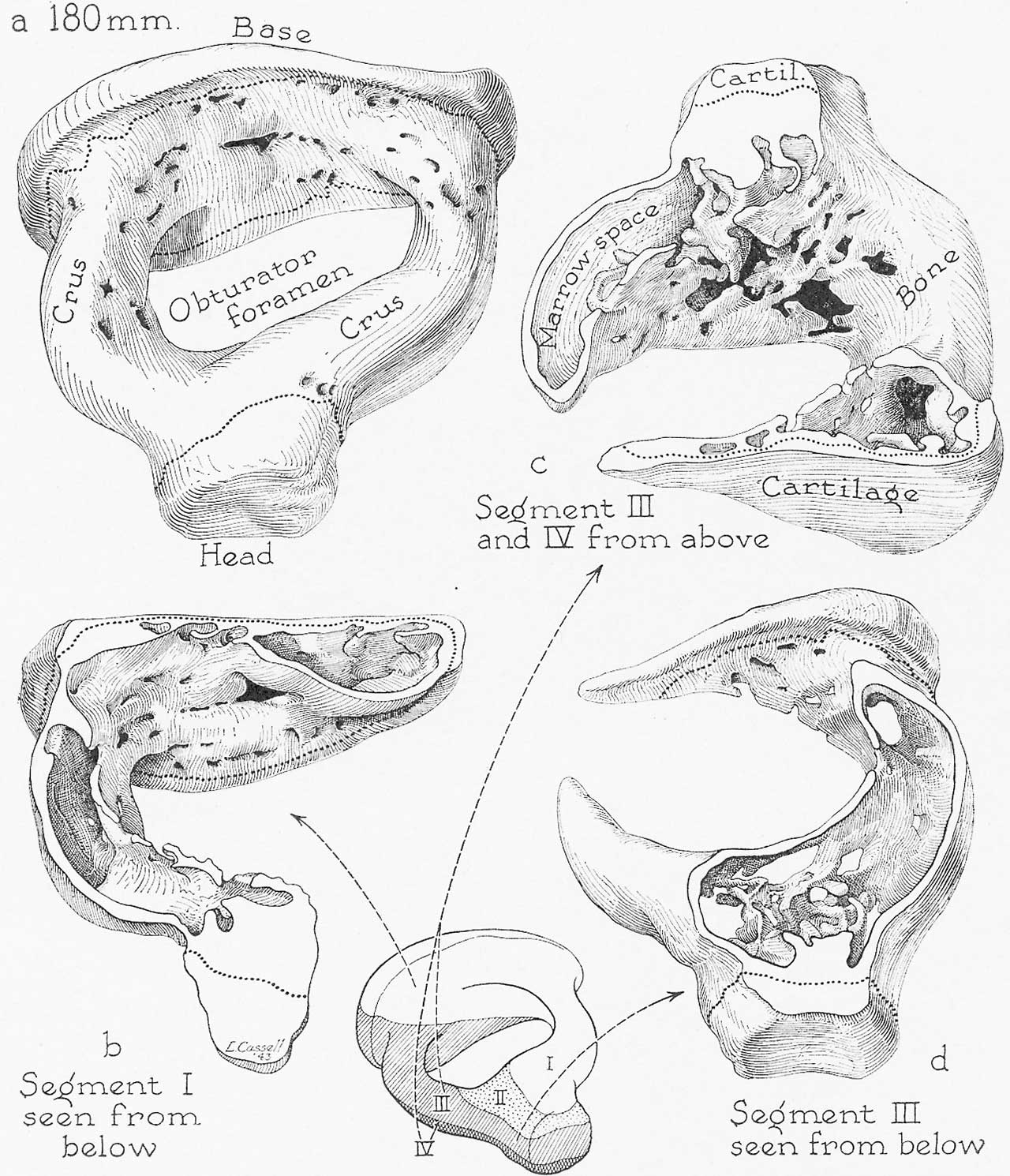 | 1,280 × 1,490 (289 KB) | Z8600021 (talk | contribs) | |
| 20:50, 16 October 2017 | 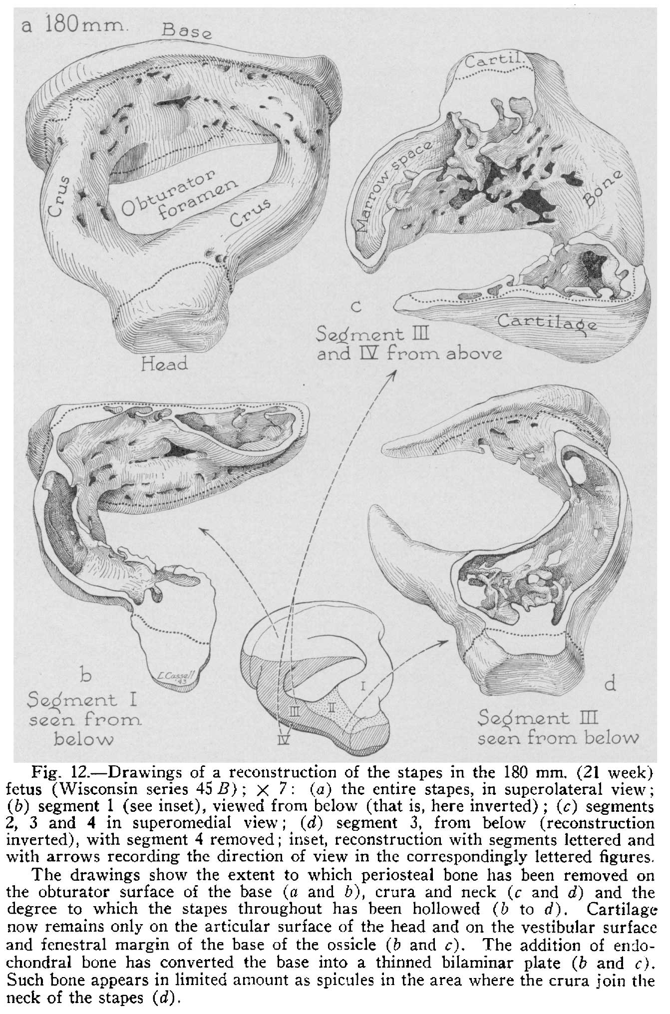 | 1,342 × 2,031 (380 KB) | Z8600021 (talk | contribs) |
You cannot overwrite this file.
File usage
The following 2 pages use this file: