File:Anson1948 fig03.jpg: Difference between revisions
(===Reference=== {{Ref-Anson1948}} {{Footer}} Category:Middle EarCategory:Historic EmbryologyCategory:1940's) |
mNo edit summary |
||
| Line 1: | Line 1: | ||
==Fig. 3. Drawings of the stapes and the adjacent fissular region of the otic capsule== | |||
Drawings (semi-diagrammatic) from Edinger tracings of the stapes and the adjacent fissular region of the otic capsule, showing developmental changes in the ossicle and in the fissular part of the capsule; X 6.6. Further developmental steps are recorded in the succeeding five plates of figures. In this, and in the five following plates, regular stippling represents unaltered cartilage; less dense stippling stands for rarefied cartilage; the areas treated in black represent bone. Parts a to c are from a 167 mm. (20 weeks) fetus (Wisconsin series 105) ; (a) slide 23, section 8; (b) slide 21, section 9; (C) slide 19, section 9). Part d is from a 163 mm. (19 week) fetus (Wisconsin series 33; slide 17, section 8). Here, a is taken at the tympanic (cranial. or superior) orifice of the fissula ante fenestram; b, at the fenestral (intermediate) orifice; c, at the vestibular (caudal, or inferior) orifice and throifigh the anterior crus of the stapes, and d, through the body, or midportion, of the fissula. | |||
Abbreviations in this and in succeeding plates are interpreted as follows: Am‘. or Ant. ems, anterior crus; Cartif, cartilage; fen. orif., fenestral orifice (of fissula ante fenestram); Post. or Post. crus, posterior crus; Tymp. Ca?,’., or Tjmzp. cavity, tympanic cavity (middle ear); tymp. 0rif., tympanic orifice (of fissula); V est, vestibule; vest. orifi, vestibular orifice (of fissula); V. w., vestibular (oval) window. | |||
===Reference=== | ===Reference=== | ||
{{Ref-Anson1948}} | {{Ref-Anson1948}} | ||
Revision as of 09:04, 15 October 2017
Fig. 3. Drawings of the stapes and the adjacent fissular region of the otic capsule
Drawings (semi-diagrammatic) from Edinger tracings of the stapes and the adjacent fissular region of the otic capsule, showing developmental changes in the ossicle and in the fissular part of the capsule; X 6.6. Further developmental steps are recorded in the succeeding five plates of figures. In this, and in the five following plates, regular stippling represents unaltered cartilage; less dense stippling stands for rarefied cartilage; the areas treated in black represent bone. Parts a to c are from a 167 mm. (20 weeks) fetus (Wisconsin series 105) ; (a) slide 23, section 8; (b) slide 21, section 9; (C) slide 19, section 9). Part d is from a 163 mm. (19 week) fetus (Wisconsin series 33; slide 17, section 8). Here, a is taken at the tympanic (cranial. or superior) orifice of the fissula ante fenestram; b, at the fenestral (intermediate) orifice; c, at the vestibular (caudal, or inferior) orifice and throifigh the anterior crus of the stapes, and d, through the body, or midportion, of the fissula.
Abbreviations in this and in succeeding plates are interpreted as follows: Am‘. or Ant. ems, anterior crus; Cartif, cartilage; fen. orif., fenestral orifice (of fissula ante fenestram); Post. or Post. crus, posterior crus; Tymp. Ca?,’., or Tjmzp. cavity, tympanic cavity (middle ear); tymp. 0rif., tympanic orifice (of fissula); V est, vestibule; vest. orifi, vestibular orifice (of fissula); V. w., vestibular (oval) window.
Reference
Anson BJ. and Cauldwell EW. Stapes, fissula ante fenestram and associated structures in man: V . From the fetus of 160 mm to term. (1948) 48(3): 263-300.
Cite this page: Hill, M.A. (2024, June 25) Embryology Anson1948 fig03.jpg. Retrieved from https://embryology.med.unsw.edu.au/embryology/index.php/File:Anson1948_fig03.jpg
- © Dr Mark Hill 2024, UNSW Embryology ISBN: 978 0 7334 2609 4 - UNSW CRICOS Provider Code No. 00098G
File history
Yi efo/eka'e gwa ebo wo le nyangagi wuncin ye kamina wunga tinya nan
| Gwalagizhi | Nyangagi | Dimensions | User | Comment | |
|---|---|---|---|---|---|
| current | 09:12, 15 October 2017 | 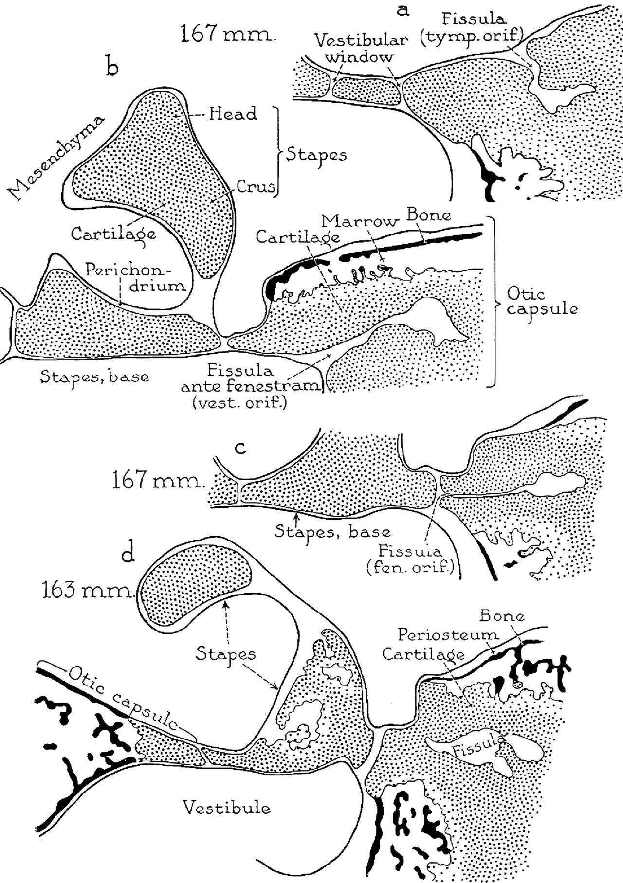 | 1,280 × 1,815 (358 KB) | Z8600021 (talk | contribs) | |
| 09:03, 15 October 2017 | 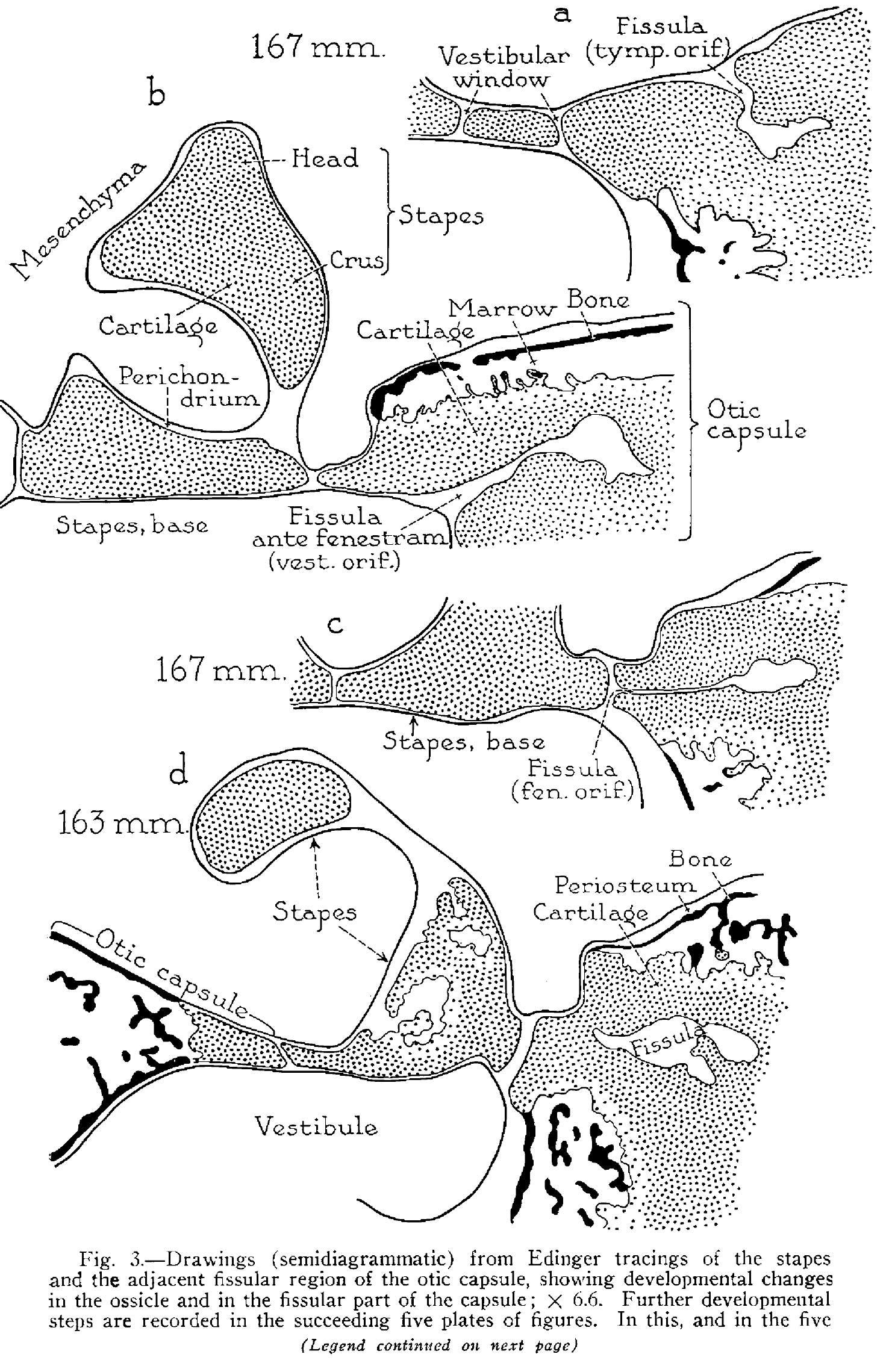 | 1,481 × 2,279 (546 KB) | Z8600021 (talk | contribs) | ===Reference=== {{Ref-Anson1948}} {{Footer}} Category:Middle EarCategory:Historic EmbryologyCategory:1940's |
You cannot overwrite this file.
File usage
The following 2 pages use this file: