File:LewisHarrison1966 fig06.jpg: Difference between revisions
From Embryology
(Fig. 6. Diagram of a reconstruction model of the embryo. A, Amniotic duct; B, collection of blood cells in yolk-sac lumen; B.S., connecting stalk; D, diverticulum of yolk-sac passing into yolk-sac duct (Y.S.D.); Ec., Ectoderm; En., endoderm; M, extra-e...) |
mNo edit summary |
||
| Line 1: | Line 1: | ||
Fig. 6. Diagram of a reconstruction model of the embryo | ==Fig. 6. Diagram of a reconstruction model of the embryo== | ||
A, Amniotic duct; B, collection of blood cells in yolk-sac lumen; B.S., connecting stalk; D, diverticulum of yolk-sac passing into yolk-sac duct (Y.S.D.); Ec., Ectoderm; En., endoderm; M, extra-embryonic mesoderm. | |||
===Reference=== | |||
{{Ref-LewisHarrison1966}} | |||
Latest revision as of 00:26, 7 August 2017
Fig. 6. Diagram of a reconstruction model of the embryo
A, Amniotic duct; B, collection of blood cells in yolk-sac lumen; B.S., connecting stalk; D, diverticulum of yolk-sac passing into yolk-sac duct (Y.S.D.); Ec., Ectoderm; En., endoderm; M, extra-embryonic mesoderm.
Reference
Lewis BV. and Harrison RG. A presomite human embryo showing a yolk-sac duct. (1966) J Anat. 100(2): 389-96. PMID: 5954785
File history
Yi efo/eka'e gwa ebo wo le nyangagi wuncin ye kamina wunga tinya nan
| Gwalagizhi | Nyangagi | Dimensions | User | Comment | |
|---|---|---|---|---|---|
| current | 00:25, 7 August 2017 | 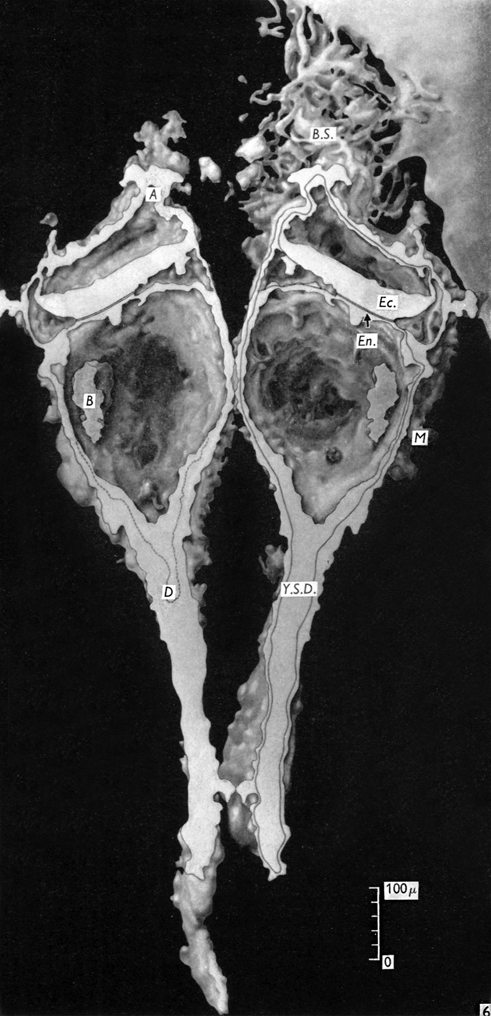 | 1,000 × 2,069 (325 KB) | Z8600021 (talk | contribs) | Fig. 6. Diagram of a reconstruction model of the embryo. A, Amniotic duct; B, collection of blood cells in yolk-sac lumen; B.S., connecting stalk; D, diverticulum of yolk-sac passing into yolk-sac duct (Y.S.D.); Ec., Ectoderm; En., endoderm; M, extra-e... |
You cannot overwrite this file.
File usage
The following page uses this file:


