File:HarrisonJeffcoate1953 plate01.jpg: Difference between revisions
(Z8600021 uploaded a new version of File:HarrisonJeffcoate1953 plate01.jpg) |
mNo edit summary |
||
| Line 1: | Line 1: | ||
==Plate 1== | ==Plate 1== | ||
Fig. 1. A low-power photomicrograph of the blastocyst (section 35). The chorionic villi are only beginning to divide, and the lacunae are well developed. The closing plug is clearly shown in the lower part of the figure. ( x 65.) | |||
Fig. 2. A section (number 27) of the anterior end of the embryo, showing the thickening of the endoderm (at a in the figure) bulging into the cavity of the yolk-sac. Since the embryo was cut obliquely, only the disc ectoderm and not the amniotic cavity is seen in this section. Part of the wall of the yolk-sac was torn during histological sectioning (just below the pointer in the figure), and was displaced into the blastocyst cavity. ( x 180.) | |||
Fig. 3. Photomicrograph of section 29, showing the amniotic cavity and the arrangement of extraembryonic mesoderm cells over its apex to simulate an amniotic duct (s.a.d.) which does not communicate with the amniotic cavity. ( x 180.) | |||
===Reference=== | ===Reference=== | ||
{{Ref-HarrisonJeffcoate1953}} | {{Ref-HarrisonJeffcoate1953}} | ||
Revision as of 21:36, 6 August 2017
Plate 1
Fig. 1. A low-power photomicrograph of the blastocyst (section 35). The chorionic villi are only beginning to divide, and the lacunae are well developed. The closing plug is clearly shown in the lower part of the figure. ( x 65.)
Fig. 2. A section (number 27) of the anterior end of the embryo, showing the thickening of the endoderm (at a in the figure) bulging into the cavity of the yolk-sac. Since the embryo was cut obliquely, only the disc ectoderm and not the amniotic cavity is seen in this section. Part of the wall of the yolk-sac was torn during histological sectioning (just below the pointer in the figure), and was displaced into the blastocyst cavity. ( x 180.)
Fig. 3. Photomicrograph of section 29, showing the amniotic cavity and the arrangement of extraembryonic mesoderm cells over its apex to simulate an amniotic duct (s.a.d.) which does not communicate with the amniotic cavity. ( x 180.)
Reference
Lewis BV. and Harrison RG. A presomite human embryo showing an early stage of the primitive streak. (1953) J Anat. 87(2):124-9. PMID: 13044724
File history
Yi efo/eka'e gwa ebo wo le nyangagi wuncin ye kamina wunga tinya nan
| Gwalagizhi | Nyangagi | Dimensions | User | Comment | |
|---|---|---|---|---|---|
| current | 21:36, 6 August 2017 | 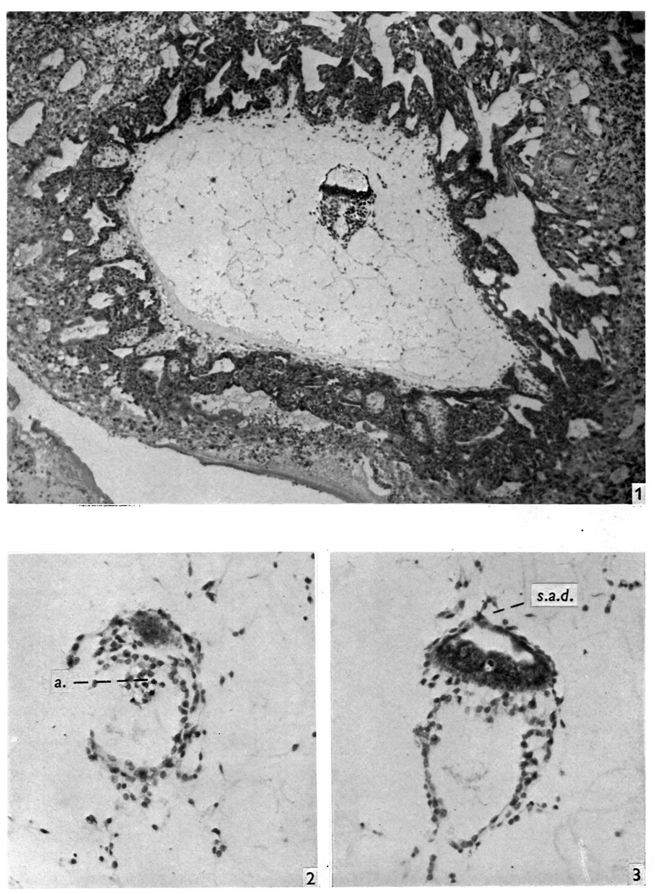 | 1,280 × 1,746 (405 KB) | Z8600021 (talk | contribs) | |
| 21:35, 6 August 2017 | 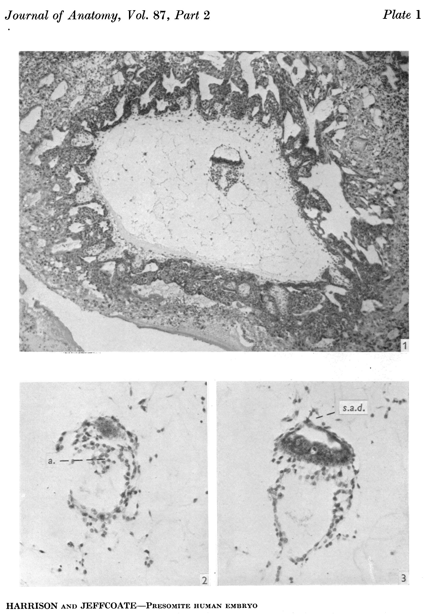 | 1,647 × 2,355 (747 KB) | Z8600021 (talk | contribs) |
You cannot overwrite this file.
File usage
The following page uses this file: