File:Kingsbury1932 plate03.jpg: Difference between revisions
mNo edit summary |
mNo edit summary |
||
| (One intermediate revision by the same user not shown) | |||
| Line 1: | Line 1: | ||
==Plate 3== | |||
16 Cat; 100-mm. fetus. Nearly median section, caudal portion of tonsil, and fascia pharyngobasilaris. The occipital bone and basisphenoid (to the right) are above. Edge of vertebra I to the left. X 16%. | |||
17 Cat; 83-mm. fetus. Nearly median section. Caudal portion of nasopharynx, cephalic aspect to the left. To show fascia pliaryiigobasilaris, basioecipital bone above, atlas and axis to the right. Tonsil not yet differentiated. Transverse pliaryngeal vein above dorsal edge of pliaryngeal musculature. X 22%. | |||
18 Kitten (125 mm., occipital crest to root of tail). Nearly median section, showing fascia pliaryiigobasilaris. Relations as above, tonsil close to skull. X 11. | |||
19 Kitten; nearly 111atu1'e (275 mm., occipital crest to root of tail). Transverse section through cephalic portion of pharyngeal tonsil. A portion of the hypopl1ysis is above; the basisphenoid with craniopliaryngeal canal underlies it. At each side of the nasopharynx the ostium tubae auditivae is shown. The soft palate is below. X 11. | |||
20 The same, but a. more caudal level. A higher nxagnlficatioii of a portion of the tonsil underlying the base of the skull. X 22%. | |||
21 Cat; young adult. Tonsil and pharyngeal epitlielimn (strati1‘ie.d and pseudostratified columnar ciliated). X 285. | |||
===Reference=== | ===Reference=== | ||
Latest revision as of 21:48, 28 March 2017
Plate 3
16 Cat; 100-mm. fetus. Nearly median section, caudal portion of tonsil, and fascia pharyngobasilaris. The occipital bone and basisphenoid (to the right) are above. Edge of vertebra I to the left. X 16%.
17 Cat; 83-mm. fetus. Nearly median section. Caudal portion of nasopharynx, cephalic aspect to the left. To show fascia pliaryiigobasilaris, basioecipital bone above, atlas and axis to the right. Tonsil not yet differentiated. Transverse pliaryngeal vein above dorsal edge of pliaryngeal musculature. X 22%.
18 Kitten (125 mm., occipital crest to root of tail). Nearly median section, showing fascia pliaryiigobasilaris. Relations as above, tonsil close to skull. X 11.
19 Kitten; nearly 111atu1'e (275 mm., occipital crest to root of tail). Transverse section through cephalic portion of pharyngeal tonsil. A portion of the hypopl1ysis is above; the basisphenoid with craniopliaryngeal canal underlies it. At each side of the nasopharynx the ostium tubae auditivae is shown. The soft palate is below. X 11.
20 The same, but a. more caudal level. A higher nxagnlficatioii of a portion of the tonsil underlying the base of the skull. X 22%.
21 Cat; young adult. Tonsil and pharyngeal epitlielimn (strati1‘ie.d and pseudostratified columnar ciliated). X 285.
Reference
Kingsbury BF. The developmental significance of the mammalian pharyngeal tonsil - Cat. (1932) Amer. J Anat. 50(2): 201-231.
Cite this page: Hill, M.A. (2024, June 26) Embryology Kingsbury1932 plate03.jpg. Retrieved from https://embryology.med.unsw.edu.au/embryology/index.php/File:Kingsbury1932_plate03.jpg
- © Dr Mark Hill 2024, UNSW Embryology ISBN: 978 0 7334 2609 4 - UNSW CRICOS Provider Code No. 00098G
File history
Yi efo/eka'e gwa ebo wo le nyangagi wuncin ye kamina wunga tinya nan
| Gwalagizhi | Nyangagi | Dimensions | User | Comment | |
|---|---|---|---|---|---|
| current | 21:46, 28 March 2017 | 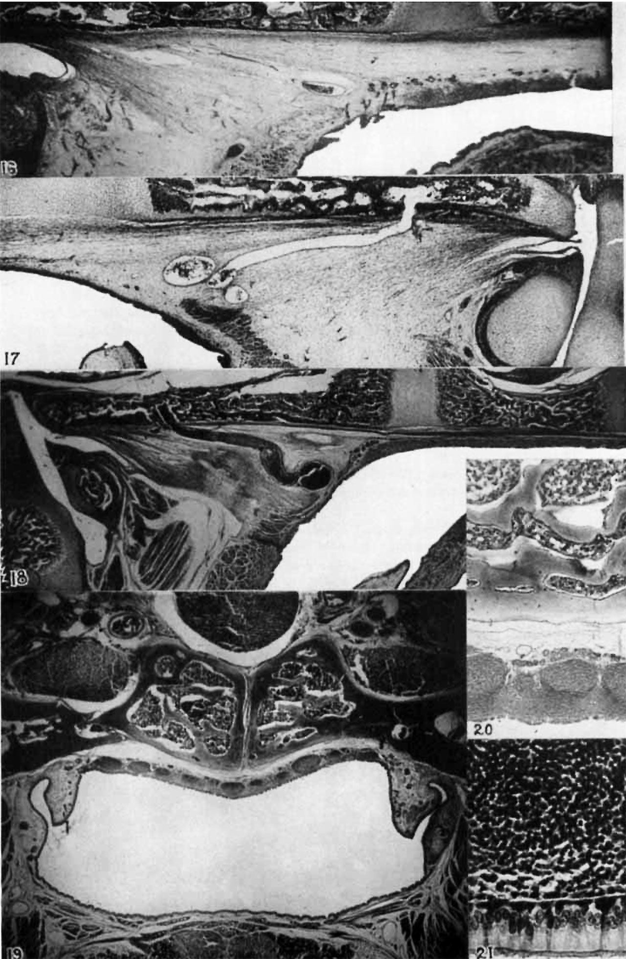 | 1,280 × 1,958 (404 KB) | Z8600021 (talk | contribs) | |
| 21:45, 28 March 2017 | 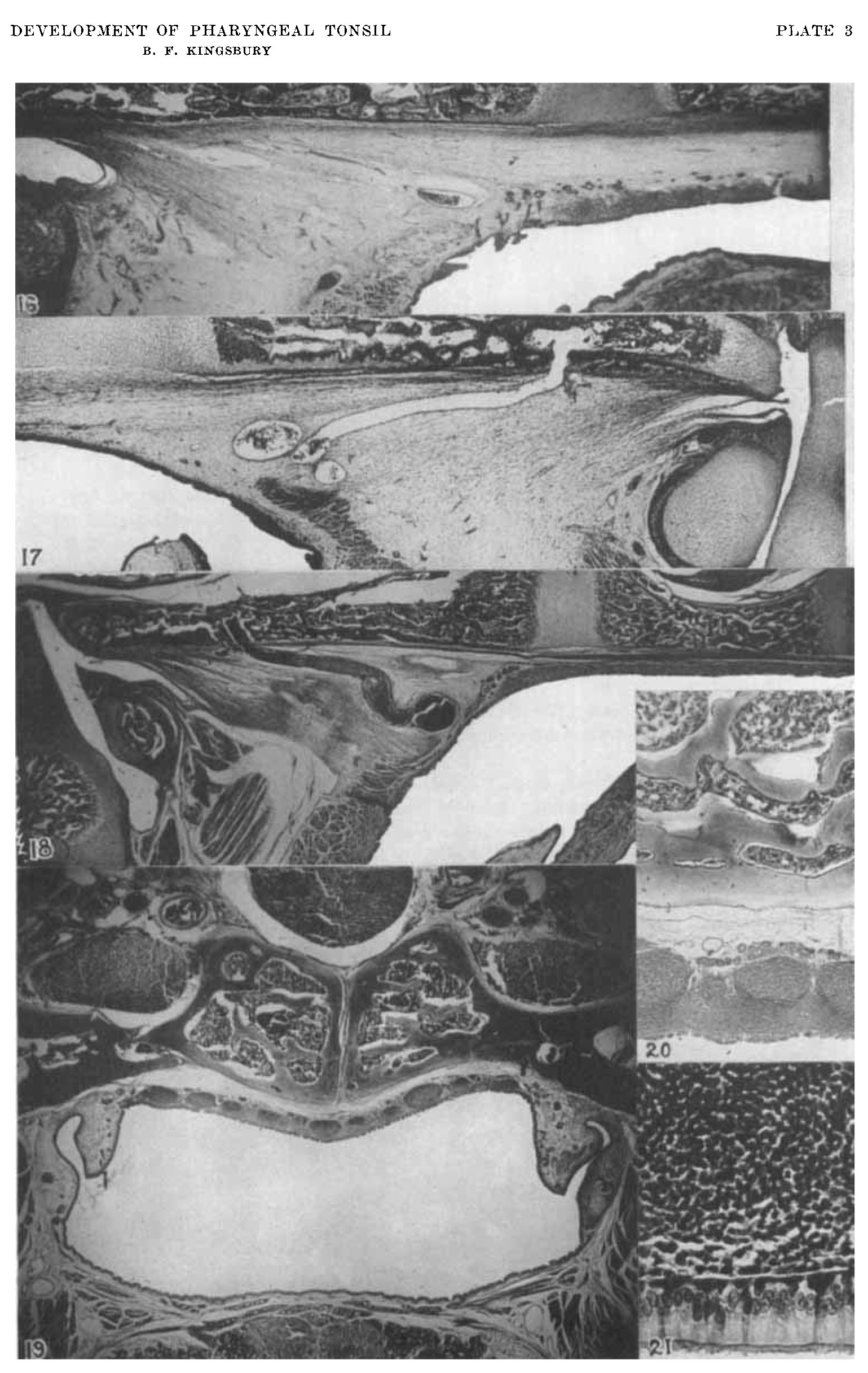 | 1,539 × 2,433 (553 KB) | Z8600021 (talk | contribs) |
You cannot overwrite this file.
File usage
The following page uses this file: