File:Odgers1935 textfig03.jpg: Difference between revisions
From Embryology
No edit summary |
mNo edit summary |
||
| (2 intermediate revisions by the same user not shown) | |||
| Line 1: | Line 1: | ||
==Text-fig. 3. Drawing of a model of the auricles in a 7.1 mm embryo== | |||
Viewed from the right side, to show the commencement of the foramen secundum. | |||
S.I. septum primum; S.s. septum spurium; F.I. foramen primum; F.II. foramen secundum; R.V.V. right venous valve. | |||
===Reference=== | |||
{{Ref-Odgers1935}} | |||
{{Footer}} | |||
Latest revision as of 14:40, 5 March 2017
Text-fig. 3. Drawing of a model of the auricles in a 7.1 mm embryo
Viewed from the right side, to show the commencement of the foramen secundum.
S.I. septum primum; S.s. septum spurium; F.I. foramen primum; F.II. foramen secundum; R.V.V. right venous valve.
Reference
Odgers PNB. The formation of the venous valves, the foramen secundum and the septum secundum in the human heart. (1935) J. Anat., 69: 412-422. PMID 17104548
Cite this page: Hill, M.A. (2024, June 26) Embryology Odgers1935 textfig03.jpg. Retrieved from https://embryology.med.unsw.edu.au/embryology/index.php/File:Odgers1935_textfig03.jpg
- © Dr Mark Hill 2024, UNSW Embryology ISBN: 978 0 7334 2609 4 - UNSW CRICOS Provider Code No. 00098G
File history
Yi efo/eka'e gwa ebo wo le nyangagi wuncin ye kamina wunga tinya nan
| Gwalagizhi | Nyangagi | Dimensions | User | Comment | |
|---|---|---|---|---|---|
| current | 14:38, 5 March 2017 |  | 585 × 800 (98 KB) | Z8600021 (talk | contribs) | |
| 14:38, 5 March 2017 | 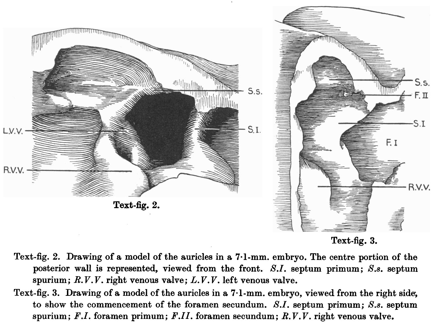 | 1,545 × 1,145 (315 KB) | Z8600021 (talk | contribs) |
You cannot overwrite this file.
File usage
The following page uses this file: