File:Anderson2016-fig01.jpg: Difference between revisions
From Embryology
| Line 1: | Line 1: | ||
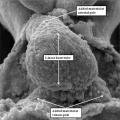
==Fig. 1. Mouse E8 linear heart tube (SEM)== | ==Fig. 1. Mouse E8 linear heart tube (SEM)== | ||
The image is a scanning electron micrograph showing the linear heart tube of the mouse within the pericardial cavity. | The image is a scanning electron micrograph showing the linear heart tube of the mouse ([[:Category:Mouse E8.0|E8]]) within the pericardial cavity. | ||
===Reference=== | ===Reference=== | ||
| Line 7: | Line 7: | ||
{{Footer}} | {{Footer}} | ||
[[Category:Mouse E8.0]][[Category:Mouse]][[Category:Scanning EM]] | |||
Revision as of 13:50, 16 February 2017
Fig. 1. Mouse E8 linear heart tube (SEM)
The image is a scanning electron micrograph showing the linear heart tube of the mouse (E8) within the pericardial cavity.
Reference
Anderson RH. Teratogenecity in the setting of cardiac development and maldevelopment. (2016)
Cite this page: Hill, M.A. (2024, June 16) Embryology Anderson2016-fig01.jpg. Retrieved from https://embryology.med.unsw.edu.au/embryology/index.php/File:Anderson2016-fig01.jpg
- © Dr Mark Hill 2024, UNSW Embryology ISBN: 978 0 7334 2609 4 - UNSW CRICOS Provider Code No. 00098G
File history
Click on a date/time to view the file as it appeared at that time.
| Date/Time | Thumbnail | Dimensions | User | Comment | |
|---|---|---|---|---|---|
| current | 13:47, 16 February 2017 |  | 800 × 800 (93 KB) | Z8600021 (talk | contribs) | ===Reference=== {{Ref-Anderson2016}} {{Footer}} |
You cannot overwrite this file.
File usage
The following 3 pages use this file: