File:Whitehead1904 fig01.jpg: Difference between revisions
From Embryology
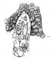
(Fig. 1. Pig 22, mm. Shows the structure of the lntertubular tisue. Mallory’s connective—tissue stain. x 800.) |
(No difference)
|
Revision as of 09:09, 24 January 2017
Fig. 1. Pig 22, mm. Shows the structure of the lntertubular tisue. Mallory’s connective—tissue stain. x 800.
File history
Yi efo/eka'e gwa ebo wo le nyangagi wuncin ye kamina wunga tinya nan
| Gwalagizhi | Nyangagi | Dimensions | User | Comment | |
|---|---|---|---|---|---|
| current | 09:09, 24 January 2017 |  | 800 × 881 (90 KB) | Z8600021 (talk | contribs) | |
| 09:09, 24 January 2017 |  | 1,342 × 1,086 (142 KB) | Z8600021 (talk | contribs) | Fig. 1. Pig 22, mm. Shows the structure of the lntertubular tisue. Mallory’s connective—tissue stain. x 800. |
You cannot overwrite this file.
File usage
The following page uses this file: