File:Atwell1918 fig24.jpg: Difference between revisions
mNo edit summary |
mNo edit summary |
||
| Line 1: | Line 1: | ||
==Fig. 24, Model of hypophysis right side from nineteen-day embryo== | |||
X 50. Brain wall, sphenoid cartilage and nasal epithelium not reconstructed. Figure 24, from right side; figure 25, dorsally and from right side; figure 26, dorsally. c.h. and 71.71., caudal and nasal horns, of pars tuberalis; 712., Secondary eminence. (dorsal part of ‘Mittelraum’); other abbreviations as in figure 22. | |||
{{Historic Disclaimer}} | {{Historic Disclaimer}} | ||
Latest revision as of 11:15, 12 November 2016
Fig. 24, Model of hypophysis right side from nineteen-day embryo
X 50. Brain wall, sphenoid cartilage and nasal epithelium not reconstructed. Figure 24, from right side; figure 25, dorsally and from right side; figure 26, dorsally. c.h. and 71.71., caudal and nasal horns, of pars tuberalis; 712., Secondary eminence. (dorsal part of ‘Mittelraum’); other abbreviations as in figure 22.
| Historic Disclaimer - information about historic embryology pages |
|---|
 |
See also Atwell WJ. The development of the hypophysis cerebri in man, with special reference to the pars tuberalis. (1926) Amer. J Anat. 37: 139-193.
Links: Pituitary Development | Rabbit Development
Reference
Atwell WJ. The development of the hypophysis cerebri of the rabbit (Lepus Cuniculus L.). (1918) Amer. J Anat. 24(2): 271-337
Cite this page: Hill, M.A. (2024, June 26) Embryology Atwell1918 fig24.jpg. Retrieved from https://embryology.med.unsw.edu.au/embryology/index.php/File:Atwell1918_fig24.jpg
- © Dr Mark Hill 2024, UNSW Embryology ISBN: 978 0 7334 2609 4 - UNSW CRICOS Provider Code No. 00098G
File history
Yi efo/eka'e gwa ebo wo le nyangagi wuncin ye kamina wunga tinya nan
| Gwalagizhi | Nyangagi | Dimensions | User | Comment | |
|---|---|---|---|---|---|
| current | 11:11, 12 November 2016 | 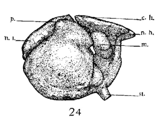 | 506 × 399 (43 KB) | Z8600021 (talk | contribs) | |
| 11:10, 12 November 2016 | 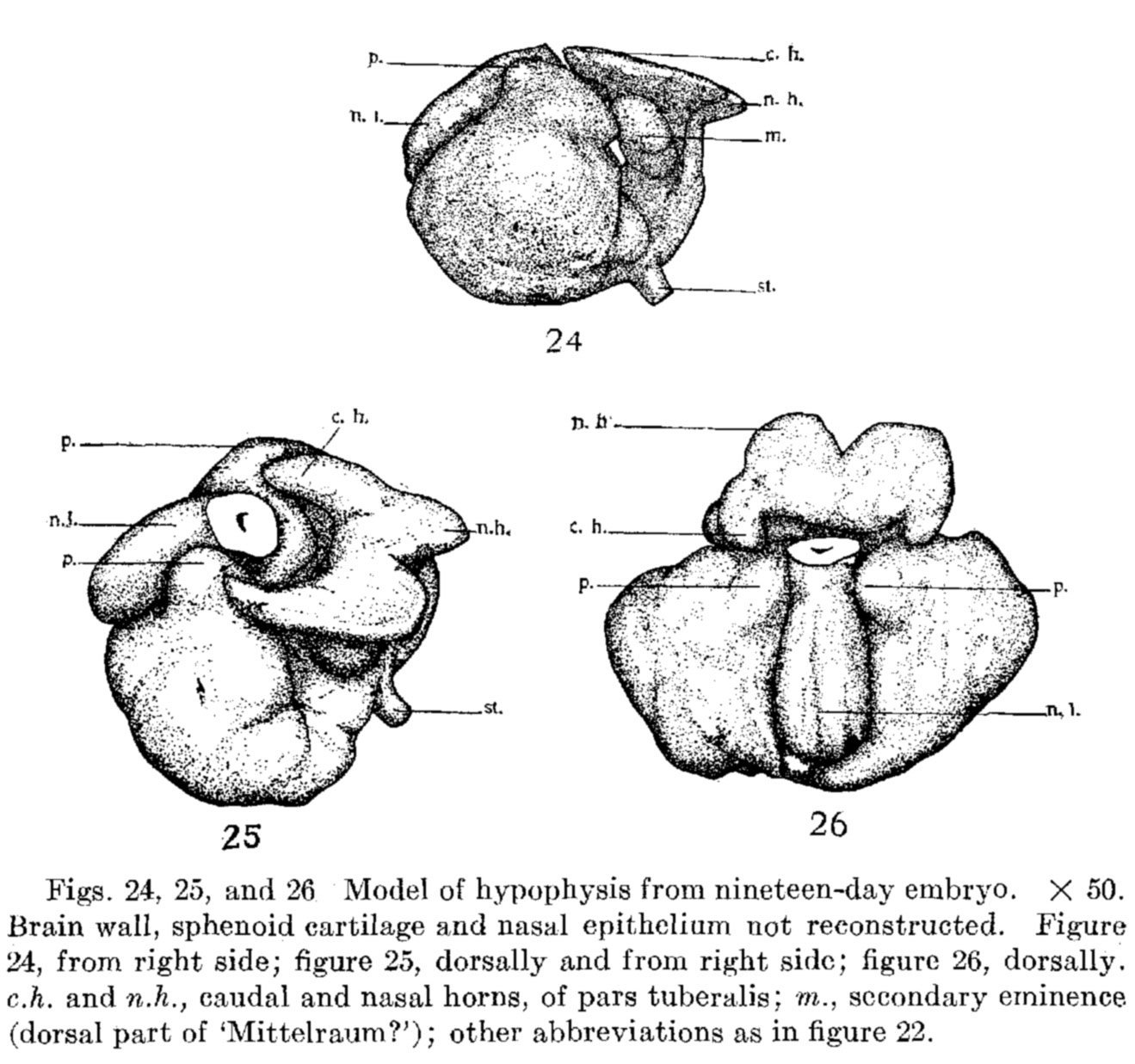 | 1,319 × 1,237 (234 KB) | Z8600021 (talk | contribs) |
You cannot overwrite this file.
File usage
The following page uses this file: