File:Senior1919 fig07.jpg: Difference between revisions
From Embryology
mNo edit summary |
mNo edit summary |
||
| Line 1: | Line 1: | ||
==Fig, 7. Indicates the course of three arteries of the embryonic lower extremity== | |||
Represented diagrammatically as they would appear if persisting in the adult. The vessels which normally persist are indicated by shading. | |||
A, A. poplitea profunda and a. interossea. | |||
B. A. peronaea posterior superficialis. d. L m. flexor digitorum longus; h, L, m. flexor hallucis longus; L p., m. tibialis posterior. | |||
===Reference=== | ===Reference=== | ||
Latest revision as of 18:42, 31 October 2016
Fig, 7. Indicates the course of three arteries of the embryonic lower extremity
Represented diagrammatically as they would appear if persisting in the adult. The vessels which normally persist are indicated by shading.
A, A. poplitea profunda and a. interossea.
B. A. peronaea posterior superficialis. d. L m. flexor digitorum longus; h, L, m. flexor hallucis longus; L p., m. tibialis posterior.
Reference
Senior HD. The development of the arteries of the human lower extremity. (1919) Amer. J Anat. 22:1-11.
Cite this page: Hill, M.A. (2024, June 19) Embryology Senior1919 fig07.jpg. Retrieved from https://embryology.med.unsw.edu.au/embryology/index.php/File:Senior1919_fig07.jpg
- © Dr Mark Hill 2024, UNSW Embryology ISBN: 978 0 7334 2609 4 - UNSW CRICOS Provider Code No. 00098G
File history
Yi efo/eka'e gwa ebo wo le nyangagi wuncin ye kamina wunga tinya nan
| Gwalagizhi | Nyangagi | Dimensions | User | Comment | |
|---|---|---|---|---|---|
| current | 18:37, 31 October 2016 | 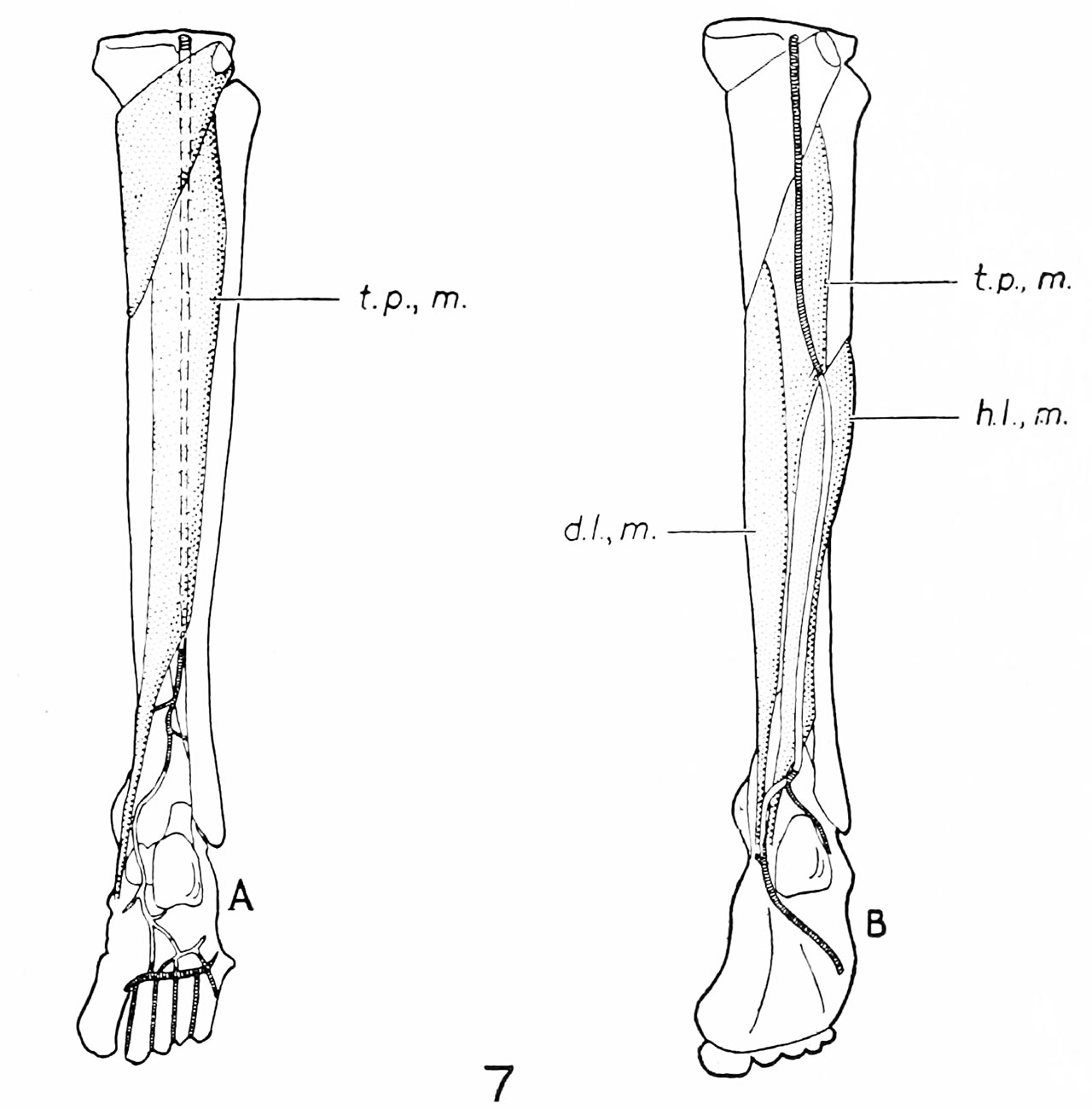 | 1,280 × 1,300 (127 KB) | Z8600021 (talk | contribs) | |
| 18:37, 31 October 2016 |  | 2,000 × 1,888 (364 KB) | Z8600021 (talk | contribs) |
You cannot overwrite this file.
File usage
The following page uses this file: