File:Ovary histology with chemotherapy.jpg: Difference between revisions
From Embryology
(Representative images of ovarian cortex before and after seven days of culture (magnification ×40), from a 15-year-old girl with lymphoma and no chemotherapy (A,B), and from a 2-year-old girl with neuroblastoma exposed to CED of 7200 mg/m2 (C,D). a) I...) |
mNo edit summary |
||
| Line 1: | Line 1: | ||
==Images of ovarian cortex before and after seven days of culture== | |||
* A, B - from a 15-year-old girl with lymphoma and no chemotherapy. | |||
* C, D - from a 2-year-old girl with neuroblastoma exposed to CED of 7200 mg/m2. | |||
a) Intact primordial follicle, b) intact secondary follicle, c) influenced primordial follicle, d) atretic follicle. | a) Intact primordial follicle, b) intact secondary follicle, c) influenced primordial follicle, d) atretic follicle. | ||
Journal.pone.0133985.g001.jpg | magnification ×40 | ||
===Reference=== | |||
<pubmed>26226487</pubmed>| [http://journals.plos.org/plosone/article?id=10.1371/journal.pone.0133985 PLoS One.] | |||
====Copyright==== | |||
© 2015 Asadi Azarbaijani et al. This is an open access article distributed under the terms of the Creative Commons Attribution License, which permits unrestricted use, distribution, and reproduction in any medium, provided the original author and source are credited. | |||
PubMed labelling added to original figure. Journal.pone.0133985.g001.jpg | |||
Revision as of 10:43, 5 September 2015
Images of ovarian cortex before and after seven days of culture
- A, B - from a 15-year-old girl with lymphoma and no chemotherapy.
- C, D - from a 2-year-old girl with neuroblastoma exposed to CED of 7200 mg/m2.
a) Intact primordial follicle, b) intact secondary follicle, c) influenced primordial follicle, d) atretic follicle.
magnification ×40
Reference
<pubmed>26226487</pubmed>| PLoS One.
Copyright
© 2015 Asadi Azarbaijani et al. This is an open access article distributed under the terms of the Creative Commons Attribution License, which permits unrestricted use, distribution, and reproduction in any medium, provided the original author and source are credited.
PubMed labelling added to original figure. Journal.pone.0133985.g001.jpg
File history
Yi efo/eka'e gwa ebo wo le nyangagi wuncin ye kamina wunga tinya nan
| Gwalagizhi | Nyangagi | Dimensions | User | Comment | |
|---|---|---|---|---|---|
| current | 10:40, 5 September 2015 | 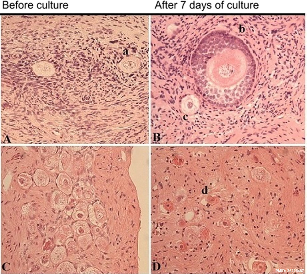 | 977 × 872 (232 KB) | Z8600021 (talk | contribs) | Representative images of ovarian cortex before and after seven days of culture (magnification ×40), from a 15-year-old girl with lymphoma and no chemotherapy (A,B), and from a 2-year-old girl with neuroblastoma exposed to CED of 7200 mg/m2 (C,D). a) I... |
You cannot overwrite this file.
File usage
The following page uses this file: