File:Streeter026.jpg: Difference between revisions
(The figures shown on this plate represent a series of median and lateral views of wax-plate reconstructions of the membranous labyrinth and the surrounding periotic tissue-spaces. They illustrate under the same scale of enlargement three typical stages in) |
mNo edit summary |
||
| (5 intermediate revisions by the same user not shown) | |||
| Line 1: | Line 1: | ||
==Fig. 26. Lateral view of a model reconstructed from a human fetus 50 mm crown-rump length== | |||
Lateral view of a model reconstructed from a human fetus 50 mm. crown-rump length (Carnegie Collection, No. 84). | |||
The cistern and the scala vestibuli are shown in green and the scala tympani is shown in orange. The scala vestibule is in the first stage of its development and consists of a row of large reticular spaces which extend from the ventral margin of the cistern downward along the apical surface of the cochlear duct. The scala tympani is more advanced and shows more complete coalescence of its constituent spaces. Enlarged 11.4 diameters. | |||
The figures shown on this [[:File:Streeter028-30.jpg|plate 4]] represent a series of median and lateral views of wax-plate reconstructions of the membranous labyrinth and the surrounding periotic tissue-spaces. They illustrate under the same scale of enlargement three typical stages in the development of these spaces. | |||
===Abbreviations=== | |||
* C. s. 1. - ductus semicircuiaris lateralis | |||
* C. s. p. - ductus semicircularis posterior | |||
* C. s. s. - ductus semicircularis superior | |||
* Duct, coch., = ductus cochlearis | |||
* Impressio rotund. - area opposite the fenestra cochleae | |||
* Impressio staped., area in contact with base of stapes | |||
* Saccus endol. - saccus endolymphaticus | |||
* Scala tymp. - scala tynipani | |||
* Scala vestib. - scala vestibule. | |||
:[[Book_-_Contributions_to_Embryology_Carnegie_Institution_No.20|'''Links''']]: [[:File:Streeter028-30.jpg|Plate 4]] | [[:File:Streeter026.jpg|Fig.26]] | [[:File:Streeter027.jpg|Fig.27]] | [[:File:Streeter028.jpg|Fig.28]] | [[:File:Streeter029.jpg|Fig.29]] | [[:File:Streeter030.jpg|Fig.30]] | [[:File:Streeter031.jpg|Fig.31]] | |||
===Reference=== | |||
Streeter G.L. '''The histogenesis and growth of the otic capsule and its contained periotic tissue-spaces in the human embryo''' Contributions to Embryology Carnegie Institution No.20 (1918) pp5-54, 4 text-figures and 4 plates. | |||
[[Category:Human]] [[Category:Historic Embryology]] [[Category:Hearing]] [[Category:George Streeter]] [[Category:Carnegie Embryo 84]] | |||
[[Category:Inner Ear]] | |||
Latest revision as of 17:31, 18 May 2015
Fig. 26. Lateral view of a model reconstructed from a human fetus 50 mm crown-rump length
Lateral view of a model reconstructed from a human fetus 50 mm. crown-rump length (Carnegie Collection, No. 84).
The cistern and the scala vestibuli are shown in green and the scala tympani is shown in orange. The scala vestibule is in the first stage of its development and consists of a row of large reticular spaces which extend from the ventral margin of the cistern downward along the apical surface of the cochlear duct. The scala tympani is more advanced and shows more complete coalescence of its constituent spaces. Enlarged 11.4 diameters.
The figures shown on this plate 4 represent a series of median and lateral views of wax-plate reconstructions of the membranous labyrinth and the surrounding periotic tissue-spaces. They illustrate under the same scale of enlargement three typical stages in the development of these spaces.
Abbreviations
- C. s. 1. - ductus semicircuiaris lateralis
- C. s. p. - ductus semicircularis posterior
- C. s. s. - ductus semicircularis superior
- Duct, coch., = ductus cochlearis
- Impressio rotund. - area opposite the fenestra cochleae
- Impressio staped., area in contact with base of stapes
- Saccus endol. - saccus endolymphaticus
- Scala tymp. - scala tynipani
- Scala vestib. - scala vestibule.
Reference
Streeter G.L. The histogenesis and growth of the otic capsule and its contained periotic tissue-spaces in the human embryo Contributions to Embryology Carnegie Institution No.20 (1918) pp5-54, 4 text-figures and 4 plates.
File history
Yi efo/eka'e gwa ebo wo le nyangagi wuncin ye kamina wunga tinya nan
| Gwalagizhi | Nyangagi | Dimensions | User | Comment | |
|---|---|---|---|---|---|
| current | 21:51, 22 April 2012 | 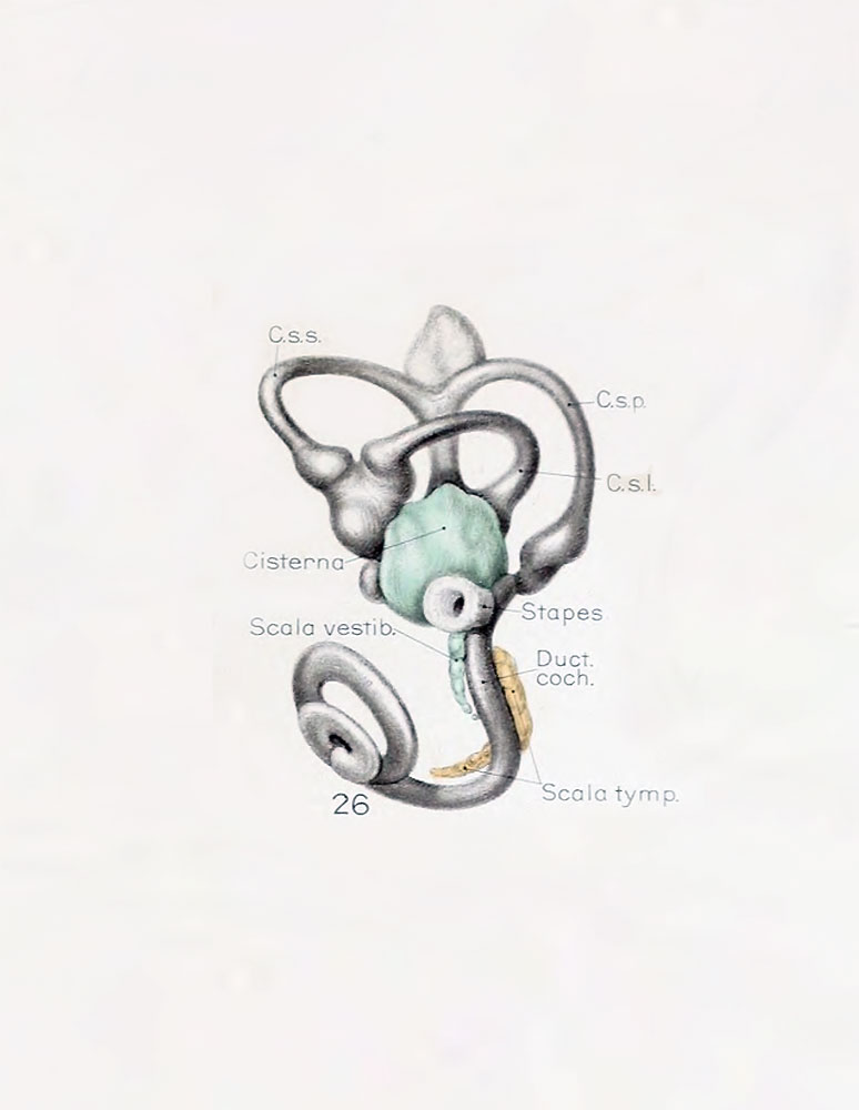 | 774 × 1,000 (45 KB) | Z8600021 (talk | contribs) | The figures shown on this plate represent a series of median and lateral views of wax-plate reconstructions of the membranous labyrinth and the surrounding periotic tissue-spaces. They illustrate under the same scale of enlargement three typical stages in |
You cannot overwrite this file.
File usage
The following 2 pages use this file: