File:Cross section of genital tubercle male.jpg: Difference between revisions
No edit summary |
mNo edit summary |
||
| Line 6: | Line 6: | ||
===Copyright=== | ===Copyright=== | ||
Beginning six months after publication, I z3417753 grant the public the non-exclusive right to copy, distribute, or display the Work under a Creative Commons Attribution-Noncommercial-Share Alike 3.0 Unported license, as described at http://creativecommons.org/licenses/by-nc-sa/3.0/ and http://creativecommons.org/licenses/by-nc-sa/3.0/legalcode. | Beginning six months after publication, I z3417753 grant the public the non-exclusive right to copy, distribute, or display the Work under a Creative Commons Attribution-Noncommercial-Share Alike 3.0 Unported license, as described at http://creativecommons.org/licenses/by-nc-sa/3.0/ and http://creativecommons.org/licenses/by-nc-sa/3.0/legalcode. | ||
--[[User:Z8600021|Mark Hill]] ([[User talk:Z8600021|talk]]) 10:21, 7 November 2014 (EST) Assessment - Student drawn image, figure relates to project topic contains only copyright and student template. There is no original source provided on which the drawing is based. File name is inaccurate. | |||
{{Template:Student Image}} | {{Template:Student Image}} | ||
Revision as of 22:50, 8 November 2014
This is a student drawn image of the cross sections of the genital tubercle in a male fetus, as it forms the tubular urethra within the penis.
References
z3417753
Copyright
Beginning six months after publication, I z3417753 grant the public the non-exclusive right to copy, distribute, or display the Work under a Creative Commons Attribution-Noncommercial-Share Alike 3.0 Unported license, as described at http://creativecommons.org/licenses/by-nc-sa/3.0/ and http://creativecommons.org/licenses/by-nc-sa/3.0/legalcode.
--Mark Hill (talk) 10:21, 7 November 2014 (EST) Assessment - Student drawn image, figure relates to project topic contains only copyright and student template. There is no original source provided on which the drawing is based. File name is inaccurate.
- Note - This image was originally uploaded as part of an undergraduate science student project and may contain inaccuracies in either description or acknowledgements. Students have been advised in writing concerning the reuse of content and may accidentally have misunderstood the original terms of use. If image reuse on this non-commercial educational site infringes your existing copyright, please contact the site editor for immediate removal.
File history
Yi efo/eka'e gwa ebo wo le nyangagi wuncin ye kamina wunga tinya nan
| Gwalagizhi | Nyangagi | Dimensions | User | Comment | |
|---|---|---|---|---|---|
| current | 20:00, 20 October 2014 | 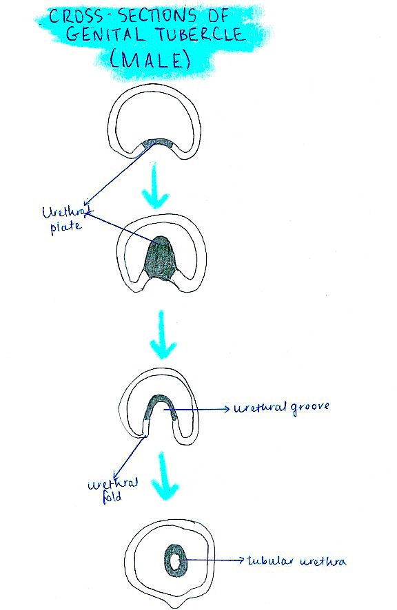 | 576 × 886 (84 KB) | Z3417753 (talk | contribs) | Reverted to version as of 09:59, 20 October 2014 |
| 20:00, 20 October 2014 |  | 576 × 886 (84 KB) | Z3417753 (talk | contribs) | Reverted to version as of 09:59, 20 October 2014 | |
| 19:59, 20 October 2014 |  | 576 × 886 (84 KB) | Z3417753 (talk | contribs) | ||
| 19:59, 20 October 2014 |  | 576 × 886 (84 KB) | Z3417753 (talk | contribs) | ||
| 19:06, 20 October 2014 | 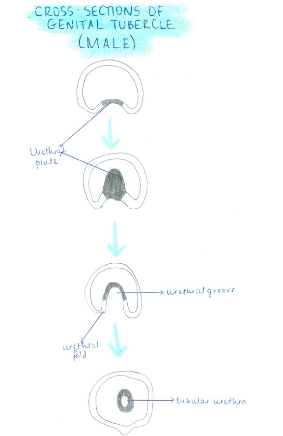 | 576 × 886 (66 KB) | Z3417753 (talk | contribs) | Cross sections of genital tubercle in a male fetus, as it forms the tubular urethra within the penis. |
You cannot overwrite this file.
File usage
The following 2 pages use this file: