File:Human fetal cochlea 01.jpg: Difference between revisions
mNo edit summary |
mNo edit summary |
||
| Line 21: | Line 21: | ||
This is an open access article distributed under the terms of the Creative Commons Attribution License (http://creativecommons.org/licenses/by/2.0), which permits unrestricted use, distribution, and reproduction in any medium, provided the original work is properly cited. | This is an open access article distributed under the terms of the Creative Commons Attribution License (http://creativecommons.org/licenses/by/2.0), which permits unrestricted use, distribution, and reproduction in any medium, provided the original work is properly cited. | ||
Figure | Figure 1. 1749-8104-8-20-1.jpg Locher et al. Neural Development 2013 8:20 doi:10.1186/1749-8104-8-20 Original figure Panel A cropped, altered in size and labelling. | ||
[[Category:Human]] [[Category:Fetal]] [[Category:Hearing]] [[Category:Inner Ear]][[Category:Week 8 | [[Category:Human]] [[Category:Fetal]] [[Category:Hearing]] [[Category:Inner Ear]][[Category:Week 8]] | ||
Revision as of 12:50, 29 June 2014
Human Fetal Cochlea (week 8.4)
(HE) staining of a cochlea at GA W10.4 (week 10 and 4 days) with higher magnification (right panel) of the basal turn cochlear duct.
Abbreviations: cd, cochlear duct; KO, Kölliker’s organ; sv, scala vestibuli; st, scala tympani.
Scale bars = 100 μm (all lower magnifications) or 50 μm (all higher magnifications).
Reference
<pubmed>24131517</pubmed>| Neural Dev.
Copyright
© 2013 Locher et al.; licensee BioMed Central Ltd. This is an open access article distributed under the terms of the Creative Commons Attribution License (http://creativecommons.org/licenses/by/2.0), which permits unrestricted use, distribution, and reproduction in any medium, provided the original work is properly cited.
Figure 1. 1749-8104-8-20-1.jpg Locher et al. Neural Development 2013 8:20 doi:10.1186/1749-8104-8-20 Original figure Panel A cropped, altered in size and labelling.
File history
Yi efo/eka'e gwa ebo wo le nyangagi wuncin ye kamina wunga tinya nan
| Gwalagizhi | Nyangagi | Dimensions | User | Comment | |
|---|---|---|---|---|---|
| current | 12:45, 29 June 2014 | 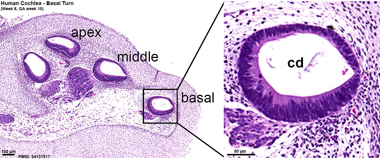 | 1,270 × 532 (266 KB) | Z8600021 (talk | contribs) | ==Human Fetal Cochlea (week 8.4)== (HE) staining of a cochlea at {{GA}} W10.4 (week 10 and 4 days) with higher magnification (right panel) of the basal turn cochlear duct. Abbreviations: cd, cochlear duct; KO, Kölliker’s organ; sv, scala vestibu... |
You cannot overwrite this file.
File usage
The following 2 pages use this file: