File:Gray0025.jpg: Difference between revisions
mNo edit summary |
|||
| Line 3: | Line 3: | ||
Diagram illustrating early formation of allantois and differentiation of body-stalk. | Diagram illustrating early formation of allantois and differentiation of body-stalk. | ||
--Mark Hill (talk) 12:38, 25 April 2013 (EST) allantois - An extraembryonic membrane, endoderm in origin extension from the early hindgut, then cloaca into the connecting stalk of placental animals, connected to the superior end of developing bladder. In reptiles and birds, acts as a reservoir for wastes and mediates gas exchange. In mammals is associated/incorporated with connecting stalk/placental cord fetal-maternal interface. | |||
{{Gray fetal membrane cartoons}} | {{Gray fetal membrane cartoons}} | ||
Revision as of 12:02, 25 February 2014
Fig. 25. Diagram illustrating early formation of allantois and differentiation of body-stalk
Diagram illustrating early formation of allantois and differentiation of body-stalk.
--Mark Hill (talk) 12:38, 25 April 2013 (EST) allantois - An extraembryonic membrane, endoderm in origin extension from the early hindgut, then cloaca into the connecting stalk of placental animals, connected to the superior end of developing bladder. In reptiles and birds, acts as a reservoir for wastes and mediates gas exchange. In mammals is associated/incorporated with connecting stalk/placental cord fetal-maternal interface.
Early embryo membrane development cartoons: Image 24 | Image 25 | Image 26 | Image 27 | Image 28
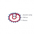
Fig 24
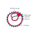
Fig 25
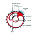
Fig 26
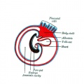
Fig 27
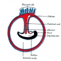
Fig 28


- Gray's Images: Development | Lymphatic | Neural | Vision | Hearing | Somatosensory | Integumentary | Respiratory | Gastrointestinal | Urogenital | Endocrine | Surface Anatomy | iBook | Historic Disclaimer
| Historic Disclaimer - information about historic embryology pages |
|---|
 |
| iBook - Gray's Embryology | |
|---|---|

|
|
Reference
Gray H. Anatomy of the human body. (1918) Philadelphia: Lea & Febiger.
Cite this page: Hill, M.A. (2024, June 26) Embryology Gray0025.jpg. Retrieved from https://embryology.med.unsw.edu.au/embryology/index.php/File:Gray0025.jpg
- © Dr Mark Hill 2024, UNSW Embryology ISBN: 978 0 7334 2609 4 - UNSW CRICOS Provider Code No. 00098G
File history
Yi efo/eka'e gwa ebo wo le nyangagi wuncin ye kamina wunga tinya nan
| Gwalagizhi | Nyangagi | Dimensions | User | Comment | |
|---|---|---|---|---|---|
| current | 11:34, 22 April 2013 |  | 500 × 500 (29 KB) | Z8600021 (talk | contribs) |
You cannot overwrite this file.
File usage
The following 15 pages use this file:
- 2009 Lecture 8
- 2010 Lecture 8
- ANAT2341 Lab 4 - Implantation and Villi Development
- ASA Meeting 2013 - Placenta
- Anatomy of the Human Body by Henry Gray
- BGDA Practical Placenta - Implantation and Early Placentation
- Coelomic Cavity Development
- Lecture - Placenta Development
- File:Gray0024-29.gif
- File:Gray0024.jpg
- File:Gray0025.jpg
- File:Gray0026.jpg
- File:Gray0027.jpg
- File:Gray0028.jpg
- Template:Gray fetal membrane cartoons