Talk:2009 Lecture 21: Difference between revisions
From Embryology
No edit summary |
No edit summary |
||
| Line 2: | Line 2: | ||
* Chemokine signaling guides regional patterning of the first embryonic artery http://genesdev.cshlp.org/content/23/19/2272.abstract?etoc | * Chemokine signaling guides regional patterning of the first embryonic artery http://genesdev.cshlp.org/content/23/19/2272.abstract?etoc | ||
:The aorta traverses the body, yet little is known about how it is patterned in different anatomical locations. Here, we show that the aorta develops from genetically distinct endothelial cells originating from diverse locations within the embryo. Furthermore, chemokine (C-X-C motif) receptor 4a (cxcr4a) is restricted to endothelial cells derived from anterior mesoderm, and is required specifically for formation of the lateral aortae. Cxcl12b, a cxcr4a ligand, is expressed in endoderm underlying the lateral aortae, and loss of cxcl12b phenocopies cxcr4a deficiency. These studies reveal unexpected endothelial diversity within the aorta that is necessary to facilitate its regional patterning by local cues. | :The aorta traverses the body, yet little is known about how it is patterned in different anatomical locations. Here, we show that the aorta develops from genetically distinct endothelial cells originating from diverse locations within the embryo. Furthermore, chemokine (C-X-C motif) receptor 4a (cxcr4a) is restricted to endothelial cells derived from anterior mesoderm, and is required specifically for formation of the lateral aortae. Cxcl12b, a cxcr4a ligand, is expressed in endoderm underlying the lateral aortae, and loss of cxcl12b phenocopies cxcr4a deficiency. These studies reveal unexpected endothelial diversity within the aorta that is necessary to facilitate its regional patterning by local cues. | ||
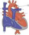
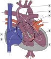
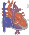
==Abnormal Heart Gallery== | |||
<gallery> | |||
Image:HeartILP_draft_aorticstenosis.jpg|Aortic Stenosis | |||
Image:HeartILP_draft_asd.jpg|ASD | |||
Image:HeartILP_draft_coarctationoftheaorta.jpg|Coarctation of the Aorta | |||
Image:HeartILP_draft_dorv.jpg|DORV | |||
Image:HeartILP_draft_funchlh.jpg|Functional Hypoplastic Left Heart | |||
Image:HeartILP_draft_hlh.jpg|Hypoplastic Left Heart | |||
Image:HeartILP_draft_interruptaorticarch.jpg|Interrupted Aortic Arch | |||
Image:HeartILP_draft_papvd.jpg|PAPVD | |||
Image:HeartILP_draft_patentda.jpg|Patent Ductus Arteriosus | |||
Image:HeartILP_draft_pulmonaryatresia.jpg|Pulmonary Atresia | |||
Image:HeartILP_draft_pulmonarystenosis.jpg|Pulmonary Stenosis | |||
Image:HeartILP_draft_tapvc.jpg|TAPVC | |||
Image:HeartILP_draft_tetralogyoffallot.jpg|Tetralogy of Fallot | |||
Image:HeartILP_draft_transposition.jpg|Transposition of the Great Vessels | |||
Image:HeartILP_draft_tricuspidatresia.jpg|Tricuspid Atresia | |||
Image:HeartILP_draft_vsd.jpg|VSD | |||
</gallery> | |||
Revision as of 13:33, 11 October 2009
Background Reading
- Chemokine signaling guides regional patterning of the first embryonic artery http://genesdev.cshlp.org/content/23/19/2272.abstract?etoc
- The aorta traverses the body, yet little is known about how it is patterned in different anatomical locations. Here, we show that the aorta develops from genetically distinct endothelial cells originating from diverse locations within the embryo. Furthermore, chemokine (C-X-C motif) receptor 4a (cxcr4a) is restricted to endothelial cells derived from anterior mesoderm, and is required specifically for formation of the lateral aortae. Cxcl12b, a cxcr4a ligand, is expressed in endoderm underlying the lateral aortae, and loss of cxcl12b phenocopies cxcr4a deficiency. These studies reveal unexpected endothelial diversity within the aorta that is necessary to facilitate its regional patterning by local cues.
Abnormal Heart Gallery
- HeartILP draft aorticstenosis.jpg
Aortic Stenosis
- HeartILP draft asd.jpg
ASD
Coarctation of the Aorta
- HeartILP draft dorv.jpg
DORV
Functional Hypoplastic Left Heart
- HeartILP draft hlh.jpg
Hypoplastic Left Heart
Interrupted Aortic Arch
- HeartILP draft papvd.jpg
PAPVD
- HeartILP draft patentda.jpg
Patent Ductus Arteriosus
- HeartILP draft pulmonaryatresia.jpg
Pulmonary Atresia
- HeartILP draft pulmonarystenosis.jpg
Pulmonary Stenosis
- HeartILP draft tapvc.jpg
TAPVC
- HeartILP draft tetralogyoffallot.jpg
Tetralogy of Fallot
- HeartILP draft transposition.jpg
Transposition of the Great Vessels
- HeartILP draft tricuspidatresia.jpg
Tricuspid Atresia
- HeartILP draft vsd.jpg
VSD


