File:Mouse bladder development E12.5-E16.5.jpg: Difference between revisions
From Embryology
(==Schematic of depicting urogenital organs adjacent to the limb and the tail at E12.5 to E16.5== A) Whole embryos from E12.5 to E16.5 B) {{HE}} sections from E12.5 to E16.5 C) Bladder developmental progression. b: bladder, bw: body wall, u: urethra...) |
mNo edit summary |
||
| Line 18: | Line 18: | ||
© 2013 Islam et al. This is an open-access article distributed under the terms of the Creative Commons Attribution License, which permits unrestricted use, distribution, and reproduction in any medium, provided the original author and source are credited. | © 2013 Islam et al. This is an open-access article distributed under the terms of the Creative Commons Attribution License, which permits unrestricted use, distribution, and reproduction in any medium, provided the original author and source are credited. | ||
Figure 1. doi:10.1371/journal.pone.0061340.g001 | Figure 1. doi:10.1371/journal.pone.0061340.g001 | ||
[[Category:Mouse]] [[Category:Mouse E12.5]] [[Category:Mouse E14.5]] [[Category:Mouse E16.5]] | [[Category:Mouse]] [[Category:Mouse E12.5]] [[Category:Mouse E14.5]] [[Category:Mouse E16.5]] | ||
Latest revision as of 01:24, 1 December 2013
Schematic of depicting urogenital organs adjacent to the limb and the tail at E12.5 to E16.5
A) Whole embryos from E12.5 to E16.5
B) (Stain - Haematoxylin Eosin) sections from E12.5 to E16.5
C) Bladder developmental progression. b: bladder, bw: body wall, u: urethra, gt: genital tubercle, up: urethral plate, r: rectuml, limb, t: tail, k: kidney.
Reference
<pubmed>23620745</pubmed>| PLoS One.
Copyright
© 2013 Islam et al. This is an open-access article distributed under the terms of the Creative Commons Attribution License, which permits unrestricted use, distribution, and reproduction in any medium, provided the original author and source are credited.
Figure 1. doi:10.1371/journal.pone.0061340.g001
File history
Yi efo/eka'e gwa ebo wo le nyangagi wuncin ye kamina wunga tinya nan
| Gwalagizhi | Nyangagi | Dimensions | User | Comment | |
|---|---|---|---|---|---|
| current | 01:23, 1 December 2013 | 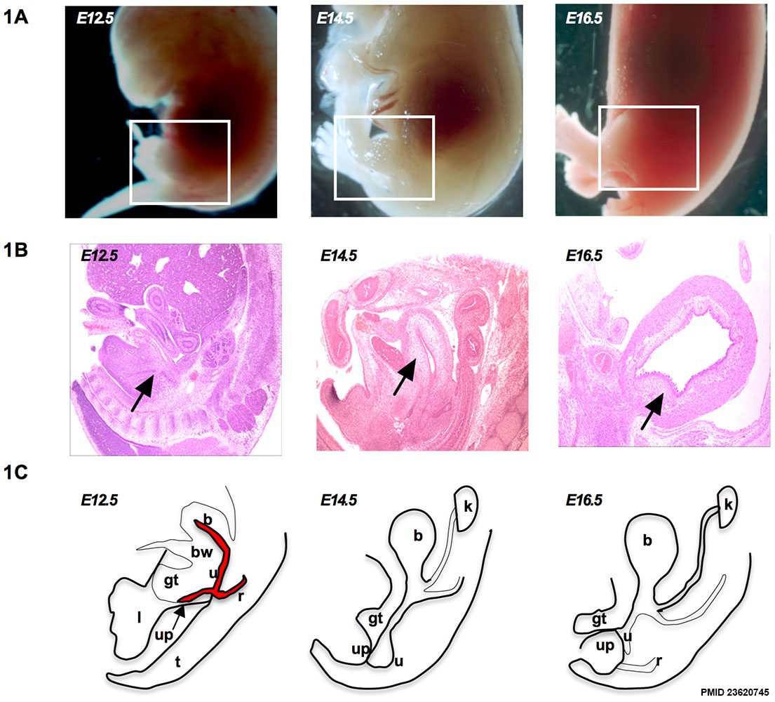 | 1,105 × 1,000 (202 KB) | Z8600021 (talk | contribs) | ==Schematic of depicting urogenital organs adjacent to the limb and the tail at E12.5 to E16.5== A) Whole embryos from E12.5 to E16.5 B) {{HE}} sections from E12.5 to E16.5 C) Bladder developmental progression. b: bladder, bw: body wall, u: urethra... |
You cannot overwrite this file.
File usage
The following page uses this file: