File:Epididymis histology 01.jpg: Difference between revisions
No edit summary |
(Z8600021 uploaded a new version of "File:Epididymis histology 01.jpg") |
(No difference)
| |
Revision as of 17:23, 27 May 2013
Epididymis Histology
Pseudostratified epithelium of the epididymis (lumen at top of image).
All cells of this type of epithelium are in contact with the basement membrane, but not all of them reach the surface of the epithelium.
Links: Histology | Histology Stains | Blue Histology images copyright Lutz Slomianka 1998-2009. The literary and artistic works on the original Blue Histology website may be reproduced, adapted, published and distributed for non-commercial purposes. See also the page Histology Stains.
Cite this page: Hill, M.A. (2024, June 26) Embryology Epididymis histology 01.jpg. Retrieved from https://embryology.med.unsw.edu.au/embryology/index.php/File:Epididymis_histology_01.jpg
- © Dr Mark Hill 2024, UNSW Embryology ISBN: 978 0 7334 2609 4 - UNSW CRICOS Provider Code No. 00098G
File history
Yi efo/eka'e gwa ebo wo le nyangagi wuncin ye kamina wunga tinya nan
| Gwalagizhi | Nyangagi | Dimensions | User | Comment | |
|---|---|---|---|---|---|
| current | 17:26, 27 May 2013 | 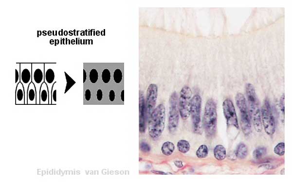 | 600 × 375 (20 KB) | Z8600021 (talk | contribs) | Reverted to version as of 04:26, 25 March 2012 |
| 17:23, 27 May 2013 |  | 400 × 534 (71 KB) | Z8600021 (talk | contribs) | ||
| 14:26, 25 March 2012 |  | 600 × 375 (20 KB) | Z8600021 (talk | contribs) | ==Epididymis Histology== |
You cannot overwrite this file.
File usage
The following page uses this file: