File:Adult human hypothalamus 01.jpg: Difference between revisions
(Z8600021 uploaded a new version of "File:Adult human hypothalamus 01.jpg") |
mNo edit summary |
||
| Line 1: | Line 1: | ||
==Adult Human Hypothalamus== | ==Adult Human Hypothalamus== | ||
3D reconstruction of the adult human hypothalamus displayed on three orthogonal | 3D reconstruction of the adult human hypothalamus displayed on three orthogonal. | ||
* '''a''' - Frontal view | |||
* '''b''' - Left lateral view | |||
* '''c''' - Inferior view | |||
T1-weighted MRI slices crossing the mammillary bodies | |||
* light blue - mammillary bodies | |||
* green plus light blue - hypothalamus | |||
* purple - anterior white commissure | |||
* brown - thalamus | |||
* purple - pallidum | |||
* yellow - third ventricle (the brain aqueduct and the ventricular foramen are also reconstructed) | |||
* dark blue - optical system | |||
===Reference=== | ===Reference=== | ||
Revision as of 18:53, 21 May 2013
Adult Human Hypothalamus
3D reconstruction of the adult human hypothalamus displayed on three orthogonal.
- a - Frontal view
- b - Left lateral view
- c - Inferior view
T1-weighted MRI slices crossing the mammillary bodies
- light blue - mammillary bodies
- green plus light blue - hypothalamus
- purple - anterior white commissure
- brown - thalamus
- purple - pallidum
- yellow - third ventricle (the brain aqueduct and the ventricular foramen are also reconstructed)
- dark blue - optical system
Reference
<pubmed>23682342</pubmed>| PMC3654779 | Surg Neurol Int.
Copyright
© 2013 Lemaire et al; This is an open-access article distributed under the terms of the Creative Commons Attribution License (http://creativecommons.org/licenses/by/2.0), which permits unrestricted use, distribution, and reproduction in any medium, provided the original work is properly cited.
SurgNeurolInt_2013_4_4_156_110667_u1.jpg
http://www.surgicalneurologyint.com/viewimage.asp?img=SurgNeurolInt_2013_4_4_156_110667_u1.jpg
File history
Click on a date/time to view the file as it appeared at that time.
| Date/Time | Thumbnail | Dimensions | User | Comment | |
|---|---|---|---|---|---|
| current | 18:38, 21 May 2013 | 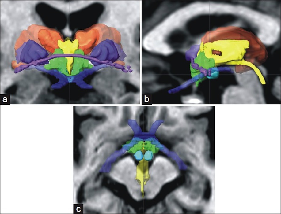 | 917 × 700 (124 KB) | Z8600021 (talk | contribs) | |
| 18:36, 21 May 2013 |  | 917 × 700 (124 KB) | Z8600021 (talk | contribs) | ==Adult Human Hypothalamus== 3D reconstruction of the adult human hypothalamus displayed on three orthogonal (a. Frontal view; b. Left lateral view; c. Inferior view) T1-weighted MRI slices crossing the mammillary bodies (light blue): hypothalamus (gre... |
You cannot overwrite this file.
File usage
There are no pages that use this file.