File:Mouse lung micro-CT 01.jpg: Difference between revisions
From Embryology
(==Mouse lung micro-CT== Representative synchrotron micro-CT image of the parenchyma of a mouse lung. The star marks a terminal bronchiole that is the terminal branch of conducting airways. Bar = 100 μm. ===Reference=== <pubmed>23533543</pubmed>...) |
mNo edit summary |
||
| Line 15: | Line 15: | ||
Figure 1 575086.fig.001.jpg | Figure 1 575086.fig.001.jpg | ||
[[Category:Mouse]] [[Category:Respiratory]] [[Category:Computed Tomography]] | |||
Latest revision as of 16:27, 10 April 2013
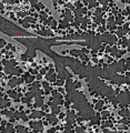
Mouse lung micro-CT
Representative synchrotron micro-CT image of the parenchyma of a mouse lung. The star marks a terminal bronchiole that is the terminal branch of conducting airways.
Bar = 100 μm.
Reference
<pubmed>23533543</pubmed>| PMC3600236 | Comput Math Methods Med
Copyright
© 2013 Luosha Xiao et al. This is an open access article distributed under the Creative Commons Attribution License, which permits unrestricted use, distribution, and reproduction in any medium, provided the original work is properly cited.
Figure 1 575086.fig.001.jpg
File history
Yi efo/eka'e gwa ebo wo le nyangagi wuncin ye kamina wunga tinya nan
| Gwalagizhi | Nyangagi | Dimensions | User | Comment | |
|---|---|---|---|---|---|
| current | 16:26, 10 April 2013 |  | 650 × 668 (113 KB) | Z8600021 (talk | contribs) | ==Mouse lung micro-CT== Representative synchrotron micro-CT image of the parenchyma of a mouse lung. The star marks a terminal bronchiole that is the terminal branch of conducting airways. Bar = 100 μm. ===Reference=== <pubmed>23533543</pubmed>... |
You cannot overwrite this file.
File usage
There are no pages that use this file.