File:Gray0613.jpg: Difference between revisions
No edit summary |
|||
| Line 1: | Line 1: | ||
==Lymphatics of | ==Lymphatics of Stomach== | ||
(Jamieson and Dobson.) | (Jamieson and Dobson.) | ||
Revision as of 23:28, 14 February 2013
Lymphatics of Stomach
(Jamieson and Dobson.)
The Gastric Glands (Figs. 613, 614) consist of two sets, superior and inferior.
Superior Gastric Glands (lymphoglandulæ gastricæ superiores) accompany the left gastric artery and are divisible into three groups, viz.: (a) upper, on the stem of the artery; (b) lower, accompanying the descending branches of the artery along the cardiac half of the lesser curvature of the stomach, between the two layers of the lesser omentum; and (c) paracardial outlying members of the gastric glands, disposed in a manner comparable to a chain of beads around the neck of the stomach (Jamieson and Dobson 110). They receive their afferents from the stomach; their efferents pass to the celiac group of preaortic glands.
Inferior Gastric Glands (lymphoglandulæ gastricæ inferiores; right gastroepiploic gland), four to seven in number, lie between the two layers of the greater omentum along the pyloric half of the greater curvature of the stomach.
Hepatic Glands (lymphoglandulæ hepaticæ) (Fig. 613), consist of the following groups: (a) hepatic, on the stem of the hepatic artery, and extending upward along the common bile duct, between the two layers of the lesser omentum, as far as the porta hepatis; the cystic gland, a member of this group, is placed near the neck of the gall-bladder; (b) subpyloric, four or five in number, in close relation to the bifurcation of the gastroduodenal artery, in the angle between the superior and descending parts of the duodenum; an outlying member of this group is sometimes found above the duodenum on the right gastric (pyloric) artery. The glands of the hepatic chain receive afferents from the stomach, duodenum, liver, gall-bladder, and pancreas; their efferents join the celiac group of preaortic glands.
(Text from Gray's Anatomy 1918)
- Gray's Images: Development | Lymphatic | Neural | Vision | Hearing | Somatosensory | Integumentary | Respiratory | Gastrointestinal | Urogenital | Endocrine | Surface Anatomy | iBook | Historic Disclaimer
| Historic Disclaimer - information about historic embryology pages |
|---|
 |
| iBook - Gray's Embryology | |
|---|---|

|
|
Reference
Gray H. Anatomy of the human body. (1918) Philadelphia: Lea & Febiger.
Cite this page: Hill, M.A. (2024, June 26) Embryology Gray0613.jpg. Retrieved from https://embryology.med.unsw.edu.au/embryology/index.php/File:Gray0613.jpg
- © Dr Mark Hill 2024, UNSW Embryology ISBN: 978 0 7334 2609 4 - UNSW CRICOS Provider Code No. 00098G
File history
Yi efo/eka'e gwa ebo wo le nyangagi wuncin ye kamina wunga tinya nan
| Gwalagizhi | Nyangagi | Dimensions | User | Comment | |
|---|---|---|---|---|---|
| current | 23:24, 14 February 2013 | 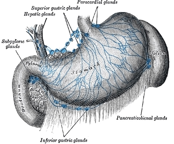 | 700 × 579 (115 KB) | Z8600021 (talk | contribs) | (Text from Gray's Anatomy 1918) {{Gray Anatomy}} Category:Immune |
You cannot overwrite this file.
File usage
The following 3 pages use this file: