File:Gray0610.jpg: Difference between revisions
((Text from Gray's Anatomy 1918) {{Gray Anatomy}} Category:Immune) |
No edit summary |
||
| Line 1: | Line 1: | ||
==The superficial lymph glands and lymphatic vessels of the lower extremity== | |||
'''Superficial Inguinal Glands''' form a chain immediately below the inguinal ligament. They receive as afferents lymphatic vessels from the integument of the penis, scrotum, perineum, buttock, and abdominal wall below the level of the umbilicus. | |||
'''Superficial Subinguinal Glands''' (lymphoglandulæ subinguinales superficiales) are placed on either side of the upper part of the great saphenous vein; their efferents consist chiefly of the superficial lymphatic vessels of the lower extremity; but they also receive some of the vessels which drain the integument of the penis, scrotum, perineum, and buttock. | |||
'''Deep Subinguinal Glands''' (lymphoglandulæ subinguinales profundæ) vary from one to three in number, and are placed under the fascia lata, on the medial side of the femoral vein. When three are present, the lowest is situated just below the junction of the great saphenous and femoral veins, the middle in the femoral canal, and the highest in the lateral part of the femoral ring. The middle one is the most inconstant of the three, but the highest, the gland of Cloquet or Rosenmüller, is also frequently absent. They receive as afferents the deep lymphatic trunks which accompany the femoral vessels, the lymphatics from the glans penis vel clitoridis, and also some of the efferents from the superficial subinguinal glands. | |||
(Text from Gray's Anatomy 1918) | (Text from Gray's Anatomy 1918) | ||
Revision as of 22:57, 14 February 2013
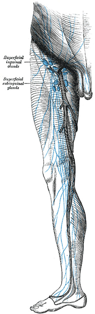
The superficial lymph glands and lymphatic vessels of the lower extremity
Superficial Inguinal Glands form a chain immediately below the inguinal ligament. They receive as afferents lymphatic vessels from the integument of the penis, scrotum, perineum, buttock, and abdominal wall below the level of the umbilicus.
Superficial Subinguinal Glands (lymphoglandulæ subinguinales superficiales) are placed on either side of the upper part of the great saphenous vein; their efferents consist chiefly of the superficial lymphatic vessels of the lower extremity; but they also receive some of the vessels which drain the integument of the penis, scrotum, perineum, and buttock.
Deep Subinguinal Glands (lymphoglandulæ subinguinales profundæ) vary from one to three in number, and are placed under the fascia lata, on the medial side of the femoral vein. When three are present, the lowest is situated just below the junction of the great saphenous and femoral veins, the middle in the femoral canal, and the highest in the lateral part of the femoral ring. The middle one is the most inconstant of the three, but the highest, the gland of Cloquet or Rosenmüller, is also frequently absent. They receive as afferents the deep lymphatic trunks which accompany the femoral vessels, the lymphatics from the glans penis vel clitoridis, and also some of the efferents from the superficial subinguinal glands.
(Text from Gray's Anatomy 1918)
- Gray's Images: Development | Lymphatic | Neural | Vision | Hearing | Somatosensory | Integumentary | Respiratory | Gastrointestinal | Urogenital | Endocrine | Surface Anatomy | iBook | Historic Disclaimer
| Historic Disclaimer - information about historic embryology pages |
|---|
 |
| iBook - Gray's Embryology | |
|---|---|

|
|
Reference
Gray H. Anatomy of the human body. (1918) Philadelphia: Lea & Febiger.
Cite this page: Hill, M.A. (2024, June 23) Embryology Gray0610.jpg. Retrieved from https://embryology.med.unsw.edu.au/embryology/index.php/File:Gray0610.jpg
- © Dr Mark Hill 2024, UNSW Embryology ISBN: 978 0 7334 2609 4 - UNSW CRICOS Provider Code No. 00098G
File history
Yi efo/eka'e gwa ebo wo le nyangagi wuncin ye kamina wunga tinya nan
| Gwalagizhi | Nyangagi | Dimensions | User | Comment | |
|---|---|---|---|---|---|
| current | 22:54, 14 February 2013 | 303 × 1,000 (81 KB) | Z8600021 (talk | contribs) | (Text from Gray's Anatomy 1918) {{Gray Anatomy}} Category:Immune |
You cannot overwrite this file.
File usage
The following 3 pages use this file: