File:Xenopus golph2 expression.jpg: Difference between revisions
(==Xenopus golph2 is expressed in epithelial cells== Stage 46/47 embryos were fixed, embedded in paraffin, sectioned to a thickness of 3 µm. Endogenous golph2 was labeled with the primary antibody 10F12, followed by anti-mouse HRP-conjugated secondary a) |
|||
| Line 2: | Line 2: | ||
Stage 46/47 embryos were fixed, embedded in paraffin, sectioned to a thickness of 3 µm. Endogenous golph2 was labeled with the primary antibody 10F12, followed by anti-mouse HRP-conjugated secondary antibody and stained with DAB solution; hematoxylin nuclear counterstaining | Stage 46/47 embryos were fixed, embedded in paraffin, sectioned to a thickness of 3 µm. Endogenous golph2 was labeled with the primary antibody 10F12, followed by anti-mouse HRP-conjugated secondary antibody and stained with DAB solution; hematoxylin nuclear counterstaining. | ||
===Legend=== | |||
{| | |||
| | |||
* '''nt''' - neural tube | |||
* '''so''' - somites | |||
* '''pn''' - pronephros | |||
* '''oe''' - esophagus | |||
* '''lu''' - lung | |||
|- | |||
| | |||
* '''lv''' - liver | |||
* '''pa''' - pancreas | |||
* '''st''' - stomach | |||
* '''i''' - intestine | |||
* '''s''' - skin | |||
|} | |||
Revision as of 13:04, 11 October 2012
Xenopus golph2 is expressed in epithelial cells
Stage 46/47 embryos were fixed, embedded in paraffin, sectioned to a thickness of 3 µm. Endogenous golph2 was labeled with the primary antibody 10F12, followed by anti-mouse HRP-conjugated secondary antibody and stained with DAB solution; hematoxylin nuclear counterstaining.
Legend
|
|
Reference
Citation: Li L, Wen L, Gong Y, Mei G, Liu J, et al. (2012) Xenopus as a Model System for the Study of GOLPH2/GP73 Function: Xenopus golph2 Is Required for Pronephros Development. PLoS ONE 7(6): e38939. doi:10.1371/journal.pone.0038939
Copyright: © 2012 Li et al. This is an open-access article distributed under the terms of the Creative Commons Attribution License, which permits unrestricted use, distribution, and reproduction in any medium, provided the original author and source are credited.
Figure 3. doi:10.1371/journal.pone.0038939.g003
File history
Yi efo/eka'e gwa ebo wo le nyangagi wuncin ye kamina wunga tinya nan
| Gwalagizhi | Nyangagi | Dimensions | User | Comment | |
|---|---|---|---|---|---|
| current | 13:01, 11 October 2012 | 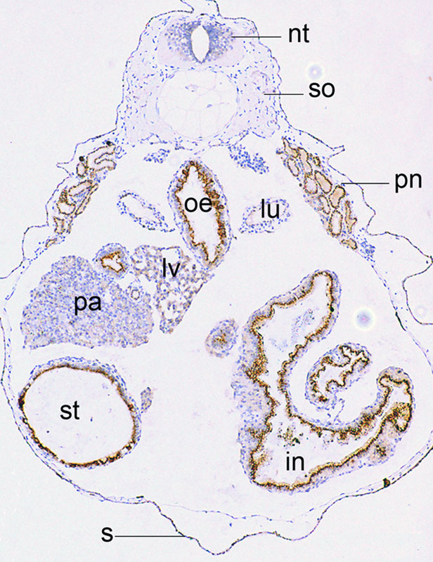 | 616 × 800 (147 KB) | Z8600021 (talk | contribs) | ==Xenopus golph2 is expressed in epithelial cells== Stage 46/47 embryos were fixed, embedded in paraffin, sectioned to a thickness of 3 µm. Endogenous golph2 was labeled with the primary antibody 10F12, followed by anti-mouse HRP-conjugated secondary a |
You cannot overwrite this file.
File usage
There are no pages that use this file.