Uploads by Z3417753
From Embryology
This special page shows all uploaded files.
| Date | Name | Thumbnail | Size | Description | Versions |
|---|---|---|---|---|---|
| 10:05, 24 October 2014 | POPs and risk of hypospadias.jpg (file) |  |
224 KB | ===Table of Maternal Serum Concentrations of PCB-153, p,p'-DDE and HCB during early pregnancy=== ====References==== <pubmed>23028613</pubmed> ====Copyright==== © Rignell-Hydbom et al. This is an open-access article distributed under the terms of the... | 1 |
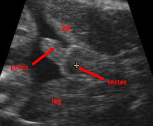
| 22:11, 23 October 2014 | Ultrasound male.jpg (file) |  |
29 KB | ===Ultrasound determining gender - boy=== Image shows an ultrasound of fetus with a penis and testes, indicating the male gender of the baby. ====Reference==== Image URL: http://upload.wikimedia.org/wikipedia/commons/a/a1/Boy.JPG Page URL:http://en... | 1 |
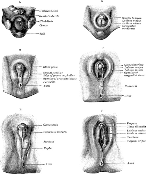
| 21:14, 23 October 2014 | Clitoris.jpg (file) |  |
102 KB | ===Stages in the Development of the External Sexual Organs in the Male and Female=== * Drawn from the Ecker-Ziegler models. ====References==== Image URL: http://commons.wikimedia.org/wiki/File:Stages_in_the_development_of_the_external_sexual_organs_i... | 1 |
| 22:33, 21 October 2014 | Schematic representation of the various treatment windows and experimental design..jpg (file) |  |
94 KB | ===Schematic representation of the various treatment windows and experimental design=== Also indicated in blue are testis differentiation in the rat (at ~embryonic day (e)13.5), reproductive tract differentiation (from ~e18.5 onwards) and the masculini... | 1 |
| 21:32, 21 October 2014 | Hypospadias.jpg (file) |  |
53 KB | ===Different types of hypospadias=== ====References==== Image URL: http://commons.wikimedia.org/wiki/File:Hypospadias-lg.jpg# Page URL: http://www.cdc.gov/ncbddd/birthdefects/hypospadias.html Original Author: Centers for Disease Control and Prevent... | 1 |
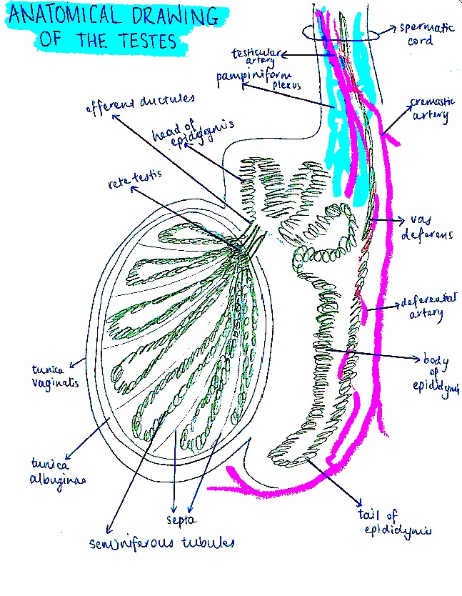
| 20:29, 20 October 2014 | Anatomical diagram of testes.jpg (file) |  |
237 KB | Reverted to version as of 10:27, 20 October 2014 | 7 |
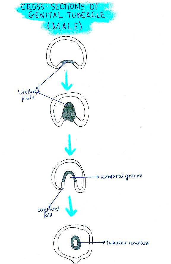
| 20:00, 20 October 2014 | Cross section of genital tubercle male.jpg (file) |  |
84 KB | Reverted to version as of 09:59, 20 October 2014 | 5 |
| 18:00, 20 October 2014 | External genitalia current model.jpg (file) |  |
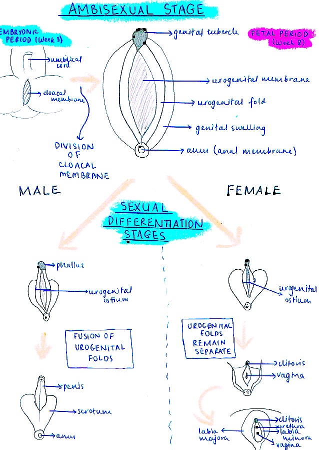
191 KB | This is a student drawn image of a flow chart of the current model describing the development of male and female external genitalia. | 1 |
| 18:04, 6 October 2014 | Flow Diagram Showing Fetal Development of External Genitalia.pdf (file) | 680 KB | 1 | ||
| 17:52, 6 October 2014 | Flow diagram of fetal development of external genitalia.pptx (file) | 755 KB | 1 | ||
| 17:49, 6 October 2014 | Flow Diagram of Fetal Development of External Genitalia.jpeg (file) |  |
65 KB | 1 | |
| 07:40, 6 October 2014 | Labelled Drawing of Testes.jpeg (file) |  |
74 KB | Anatomical drawing of the testes and epididymis | 1 |
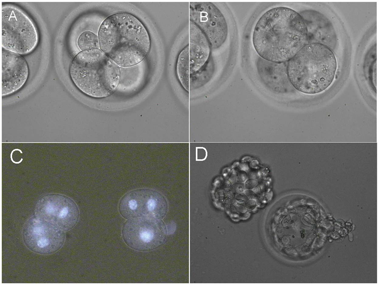
| 14:27, 19 August 2014 | Fusion of two pairs of blastomeres inside 4-cell embryos.png (file) |  |
1.39 MB | (A) Fusion of first pair of blastomeres. (B) Fusion of second pair of blastomeres. (C) Resulting 2-cell embryos. Hoechst 33342 staining confirms the presence of two nuclei in each blastomere. (D) In vitro development of 4-cell embryos with two fused pa... | 1 |